Table of Contents >> Show >> Hide
- Start Here: Your Lymphatic System (A Map You’ve Had All Along)
- What Non-Hodgkin’s Lymphoma Actually Is
- Common NHL Types You’ll Hear About (And Why Names Matter)
- Symptoms: A “Body Map” of What NHL Can Look Like
- Diagnosis: The “Detective Board” Timeline (What Usually Happens Next)
- Imaging, Explained Like a Human: CT vs PET/CT
- Staging in Plain English (The Roman Numerals You Didn’t Ask For)
- Treatment Options: The Menu (Not Everyone Orders the Same Thing)
- Side Effects and Supportive Care (The Practical Stuff That Keeps Life Livable)
- “What’s on My Pathology Report?” A Mini Decoder (With a Calm Voice)
- Questions to Ask Your Clinician (Because “Uhh… so… what now?” Is Not a Plan)
- Prognosis and Follow-Up: Reading the Road Signs Without Panic
- Real-World Experiences: What the NHL Journey Can Feel Like (500+ Words)
- Wrapping It Up
If the words “non-Hodgkin’s lymphoma” feel like they were invented to make Google searches stressful, you’re not alone.
The good news: this topic becomes a lot less intimidating once you can picture what’s happeningwhere it starts, how doctors confirm it,
and why two people with “NHL” might have very different plans.
This guide uses “visual thinking” (simple mental images, mini-diagrams, and report-decoding tips) to explain non-Hodgkin’s lymphoma in plain American English.
It’s educationalnot a diagnosis or medical advice. If something here sounds like you or someone you love, the best next step is a clinician who can look at the whole picture.
Start Here: Your Lymphatic System (A Map You’ve Had All Along)
Picture your body as a city. Your blood vessels are highways. Your lymphatic system is the city’s “drainage + security” department
moving fluid, filtering debris, and helping immune cells respond to threats.
Include labels: lymph nodes (neck, armpits, chest, abdomen, groin), lymph vessels, spleen, thymus,
bone marrow, tonsils. Add arrows showing lymph fluid moving toward the chest and back into the bloodstream.
Lymph nodes: not “bad lumps,” just tiny filters
Lymph nodes are small bean-shaped checkpoints. When your immune system is activelike during an infectionnodes can swell.
That’s why a swollen node doesn’t automatically mean cancer. The key is the pattern: how long it lasts, whether it’s growing,
and what else is happening in the body.
What Non-Hodgkin’s Lymphoma Actually Is
Non-Hodgkin’s lymphoma (NHL) is a large group of blood cancers that start in the lymphatic systemusually in
lymphocytes (a type of white blood cell). Most are B-cell lymphomas; a smaller portion are T-cell lymphomas.
One name, many “subtypes”
“NHL” is like saying “sports.” Helpful category… but it doesn’t tell you whether you’re talking about basketball or bowling.
The subtype is what drives the plan: how fast it tends to grow, which tests matter most, and which treatments are most effective.
- Indolent (slow-growing): may be monitored at first in some cases (“watch and wait” / active surveillance).
- Aggressive (faster-growing): usually needs treatment sooner, but can also be highly treatable.
- B-cell vs T-cell: different biology; different medication strategies.
Quick (respectful) contrast: Hodgkin vs non-Hodgkin
Hodgkin lymphoma is a different category with distinctive features under the microscope. Non-Hodgkin’s lymphoma includes most other lymphoma subtypes.
They can overlap in symptoms, but the diagnosis depends on tissue testing (not vibes, not guesswork, not the “shape of a lump”).
Common NHL Types You’ll Hear About (And Why Names Matter)
Diffuse Large B-Cell Lymphoma (DLBCL)
DLBCL is a common aggressive B-cell lymphoma. “Aggressive” sounds scary, but in cancer-language it often means
“grows faster” and therefore “needs treatment now,” not “no hope.” Many people respond well to standard first-line therapies.
Follicular Lymphoma
Follicular lymphoma is often slower-growing. Some people need treatment right away; others can safely be monitored for a time.
The decision depends on symptoms, organ involvement, and how the disease is behavingnot just the label.
Marginal Zone, Mantle Cell, Burkitt, and Others
These subtypes vary widely in pace and typical treatment approaches. The point isn’t to memorize every nameyour brain deserves better.
The point is: subtype = strategy.
Takeaway visual: Imagine subtype as the “operating system,” and the treatment plan as the “apps” that work best on it.
Same device category; different software requirements.
Symptoms: A “Body Map” of What NHL Can Look Like
Symptoms depend on where lymphoma is growing and how active the immune system is. Some people notice a lump; others feel “off” in a more general way.
Common symptoms (the usual suspects)
- Painless swollen lymph nodes (neck, armpit, groin are common spots you can feel).
- Unexplained fever, drenching night sweats, and unintentional weight loss (often called “B symptoms”).
- Fatigue that doesn’t match your sleep schedule.
- Itching or skin changes in some cases.
Location-based clues (because bodies love geography)
- Chest involvement: cough, shortness of breath, or pressure sensations.
- Abdomen involvement: belly swelling, feeling full quickly, discomfort.
- Bone marrow involvement: abnormal blood counts that can show up on labs.
Outline of a body silhouette with callouts: neck nodes, axillary nodes, groin nodes, spleen area (left upper abdomen),
chest nodes, fatigue icon, thermometer icon, “night sweats” icon.
A gentle reality check: infections, autoimmune issues, and even some medications can also cause swollen nodes or fatigue.
That’s why NHL diagnosis requires testingespecially a biopsy.
Diagnosis: The “Detective Board” Timeline (What Usually Happens Next)
Diagnosing NHL is less like a single test and more like assembling a puzzle: symptoms, exam findings, imaging, labs, andmost importanttissue.
Step 1: History + physical exam
Clinicians check lymph node areas, spleen/liver size, and symptom patterns. Expect questions about infections, immune conditions,
medications that suppress immunity, and family history.
Step 2: The biopsy (the “name the subtype” moment)
A biopsy takes tissue from an enlarged node or affected area so a pathologist can look at cell patterns and run lab markers.
This is how doctors confirm lymphoma and identify the subtype. Blood tests alone can’t do that job reliably.
Step 3: Lab work and specialized testing
You may hear terms like immunohistochemistry or flow cytometrythese help identify which proteins are on the cells
(often crucial for classification and treatment selection). Blood tests can also evaluate blood counts, organ function, and sometimes “tumor activity” clues.
Step 4: Imaging (seeing the full map)
Imaging helps show where lymphoma is and how extensive it is. The most common tools are CT and PET/CT, depending on subtype and clinical context.
Flow: Symptoms / lump → exam + labs → imaging → biopsy → subtype confirmed → staging → treatment plan
Note: Biopsy is the hinge point where “maybe” becomes “this specific subtype.”
Sometimes: bone marrow biopsy or spinal fluid testing
Not everyone needs these. They’re used in specific situations based on subtype, symptoms, or imaging/lab findings.
If they’re recommended, it’s usually because the results could meaningfully change staging or treatment.
Imaging, Explained Like a Human: CT vs PET/CT
CT (Computed Tomography)
Think of a CT scan as a high-detail “anatomy photo set.” It can show enlarged lymph nodes or organs and help measure them over time.
PET/CT
PET adds a “metabolic activity” layer. Many lymphomas take up a small amount of a sugar-like tracer (often FDG), so active areas can light up.
PET/CT combines the metabolic map (PET) with the anatomy map (CT), which can be useful for staging and for checking response to treatment in many cases.
- CT: shape + size (“what it looks like”)
- PET: activity (“how busy it is”)
- PET/CT: both layers aligned (“busy spots, pinned to exact locations”)
How to read a scan report without spiraling
Scan reports often list every findingimportant, unimportant, and “probably nothing but noted anyway.”
Helpful strategy: focus on (1) the impression section, and (2) what your clinician says it means for staging or next steps.
Staging in Plain English (The Roman Numerals You Didn’t Ask For)
Staging describes where lymphoma is in the body and how widespread it is. For many adult NHL subtypes,
clinicians often use the Lugano/Ann Arbor-style staging framework (Stages I–IV), plus a few add-ons.
The simple mental picture
- Stage I: one lymph node region (or one nearby lymphoid area).
- Stage II: two or more lymph node regions on the same side of the diaphragm (all above or all below).
- Stage III: lymph node regions on both sides of the diaphragm.
- Stage IV: more diffuse involvement of organs like bone marrow, liver, lungs, etc. (depends on subtype and context).
Extra letters and words you might see
- E: extranodal extension (involvement outside lymph nodes).
- Bulky: a larger mass (definitions vary by guideline/subtype).
- B symptoms: fever/night sweats/weight loss clusterclinically meaningful for many cases.
Important nuance: in lymphoma, higher stage does not automatically mean “untreatable.”
Some advanced-stage lymphomas respond very well to therapy, while some early-stage indolent lymphomas can behave quietly for years.
Draw a simple torso with a horizontal “diaphragm” line. Color nodes above vs below. Show stage II as “same side,” stage III as “both sides.”
Treatment Options: The Menu (Not Everyone Orders the Same Thing)
Treatment depends on subtype, stage, symptoms, overall health, and how quickly the lymphoma is progressing. Your care team’s goal is to choose
the most effective approach with the best risk-benefit balance for your situation.
Active surveillance (“watch and wait”)
For some slower-growing lymphomas without symptoms or organ risk, the safest move can be monitoring with scheduled visits and tests.
It’s not “doing nothing.” It’s “not treating before treatment is needed.”
Chemo-immunotherapy (a common backbone for many B-cell NHLs)
Many regimens combine chemotherapy with targeted immune-based drugs such as monoclonal antibodies.
One widely used example for certain aggressive B-cell lymphomas is a chemo combination plus an anti-CD20 antibody (often recognized by patients as “R-CHOP”).
The exact plan and number of cycles vary by subtype and stage, so this is always individualized.
Targeted therapy
Targeted therapies aim at specific pathways cancer cells rely on. Depending on subtype, these can include oral medications, antibody-drug combinations,
or other targeted agents. The “target” is determined by the biology of the lymphoma and prior treatments.
Radiation therapy
Radiation may be used for localized disease in some cases, for symptom relief, or as part of a combined approach. It’s very “aimed,” not full-body.
Cellular therapy and transplant (for specific scenarios)
For some relapsed or refractory lymphomas, advanced options like CAR T-cell therapy or stem cell transplant
may be discussed. These are specialized treatments typically offered at centers with relevant expertise.
Start: subtype → (indolent vs aggressive) → symptoms? organ risk? stage? → surveillance vs therapy.
Add a branch for “relapsed/refractory” → clinical trials / CAR T / transplant discussions.
Side Effects and Supportive Care (The Practical Stuff That Keeps Life Livable)
Side effects vary by treatment type and by person. Many are manageable, and your team wants to know about them earlybefore “minor” becomes “miserable.”
Common themes
- Fatigue: common during and after treatment; pacing helps more than “pushing through.”
- Lower blood counts: can increase infection risk or cause anemia-related tiredness.
- Nausea, appetite changes, GI upset: often treatable with supportive medications and food strategies.
- Nerve symptoms: some drugs can cause tingling or numbness; report early.
Practical safety reminders
If treatment affects immune function, your care team may recommend vaccine timing, infection-prevention steps, and what symptoms should trigger a call.
Don’t guessget the clinic’s “if X happens, do Y” instructions in writing.
“What’s on My Pathology Report?” A Mini Decoder (With a Calm Voice)
Pathology reports can look like they were written by three specialists and a keyboard cat. Here are common components and what they’re for:
Key items you might see
- Subtype name: the diagnosis (for example, “diffuse large B-cell lymphoma”).
- Immunophenotype/markers: proteins on the cell surface used to classify the lymphoma (and sometimes guide therapy).
- Grade or proliferation notes: clues about growth behavior in certain subtypes.
- Genetic/cytogenetic testing: identifies specific changes that can matter for risk and treatment decisions in some cases.
Show a mock report with labeled boxes: Diagnosis, Microscopic description, Immunostains/flow, Genetics, Comment/summary.
Add a sticky note: “Ask: What subtype is it? What stage? What’s the goal of treatment?”
Questions to Ask Your Clinician (Because “Uhh… so… what now?” Is Not a Plan)
- What exact subtype is this, and how does it typically behave?
- What stage is it, and what tests were used to determine that?
- Is treatment needed now, or is active surveillance appropriate?
- What’s the goal: cure, long-term control, symptom relief, or something else?
- What side effects should we watch for, and which ones are urgent?
- Should I consider a second opinion or a lymphoma specialty center?
- Are clinical trials relevant for my subtype/stage?
Prognosis and Follow-Up: Reading the Road Signs Without Panic
Prognosis in NHL depends heavily on subtype, stage, and individual factors (like overall health and certain lab results).
For some lymphomas, “stage” is only part of the storybiology can matter just as much.
Follow-up often includes clinic visits, lab work, and sometimes imaging, especially in the first years after treatment
when clinicians are tracking response and watching for recurrence or late effects. Your team may also discuss survivorship care:
fatigue recovery, heart health, vaccinations, and emotional support resources.
And yesscan anxiety is real. (If it had a frequent-flyer program, many patients would qualify for platinum status.)
The goal is to build a follow-up plan that is medically appropriate and emotionally survivable.
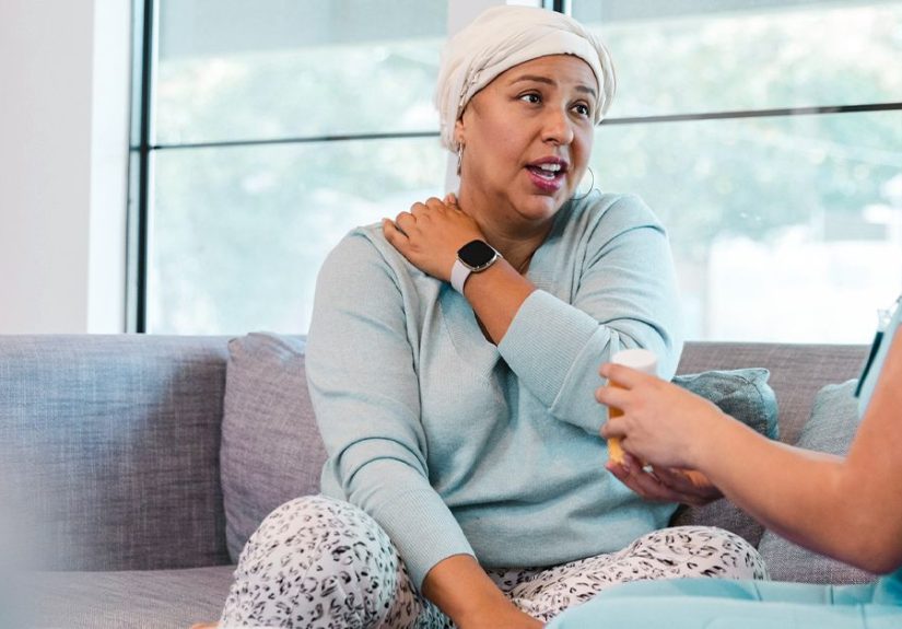
Real-World Experiences: What the NHL Journey Can Feel Like (500+ Words)
The most “visual” part of non-Hodgkin’s lymphoma isn’t always the scanit’s the day-to-day experience of uncertainty turning into a plan.
Many people describe the early phase as a strange mix of not that sick and not that okay: a lump that doesn’t hurt,
fatigue that doesn’t match the calendar, or night sweats that make the laundry basket feel personally offended.
A common experience is the “two-speed world” effect. Life around you keeps moving at normal speedschool drop-offs, work emails, dinner plans
while your own timeline suddenly becomes measured in appointments: first a primary care visit, then a specialist, then imaging, then the biopsy.
People often say the waiting is harder than the testing. A scan takes minutes; waiting for the result can feel like your brain is running
a marathon in flip-flops.
When the diagnosis arrives, the subtype name can feel like a new language you didn’t sign up to learn. Many patients and caregivers describe a mental
pivot point: the moment they realize “NHL” isn’t one disease, and that knowing the subtype is like finally getting the right map.
Before that, it can feel like being told, “You’re going on a trip,” without being told whether you need a swimsuit or a snow shovel.
During treatmentif treatment is neededpeople often find that the emotional rhythm changes. The first cycle can be the most intimidating because everything is unknown:
How tired will I feel? Will I be nauseated? What happens if I spike a fever? Over time, many people become surprisingly skilled at “symptom pattern recognition,”
learning what’s normal for them and what deserves a call. Caregivers frequently describe their own job as part scheduler, part encourager, part note-taker,
and part snack negotiator (“Yes, soup counts as a meal; no, ice cream is not a vegetable.”).
For those on active surveillance, the experience is different but still real: the awkward space of having cancer and not “doing something” immediately.
Many patients say it helps to rename it in their minds as active monitoringbecause it is active. It’s data gathering. It’s protection against overtreatment.
Still, it can bring “appointment dread” and a special kind of anxiety that spikes before each follow-up visit.
Across many stories, a few practical coping strategies show up again and again:
writing questions down before appointments (because stress deletes memory like it’s doing spring cleaning),
bringing a second person to listen, asking for a clear “if this, then that” action plan, and finding one reliable place to keep results and notes.
People also often mention that support can come from unexpected placesfriends who are good at logistics, not speeches; communities that understand “scanxiety” without explanation;
and clinicians who take five extra minutes to translate the medical language into human language.
The most hopeful theme is also the most grounded: once the diagnosis is clear and the plan is in motion, many people feel less afraid than they did in the “maybe” phase.
Non-Hodgkin’s lymphoma is serious, but it’s also a field where classification and treatment options have expanded dramaticallyand where individualized plans are the norm.
The journey is rarely linear, but it is very often navigable, especially with a team that treats both the disease and the person carrying it.