Table of Contents >> Show >> Hide
- White matter vs gray matter: a quick brain map (no pop quiz)
- What MS is doing under the hood
- How MS affects white matter
- How MS affects gray matter (the part people forget to invite to the conversation)
- How white matter and gray matter damage are connected
- What doctors look for: MRI clues in white and gray matter
- Why disability can progress even when white matter lesions don’t “look worse”
- Can treatment protect white and gray matter?
- Specific examples: how white and gray matter changes can show up day-to-day
- When to contact a clinician
- Conclusion: the big takeaways
- Experiences: What living with white- and gray-matter changes can be like
Multiple sclerosis (MS) has a reputation for leaving “spots” on brain scansthose classic white matter lesions that look like tiny constellations on MRI.
But here’s the plot twist: MS doesn’t limit itself to white matter highways. It can also damage the brain’s gray matter “neighborhoods,” where nerve cell
bodies and processing centers live. And that matters, because gray matter injury is strongly tied to symptoms people actually noticelike slowed thinking,
fatigue, mood changes, and physical disability.
In this article, we’ll break down what white matter and gray matter do, how MS can disrupt both, what doctors look for on MRI, and why two people with
“similar-looking” scans can feel very different. You’ll also get real-world examples, practical context, and a 500-word experiences section at the end
to round things out.
White matter vs gray matter: a quick brain map (no pop quiz)
White matter: the brain’s wiring and long-distance routes
White matter is mostly made of axonslong nerve fibers that carry electrical signals from one brain region to another. Many axons are wrapped in myelin,
a fatty insulating layer that helps signals travel quickly and efficiently (think: insulation around electrical wires). When myelin is damaged, signal
speed drops, signals get scrambled, or messages fail to arrive on time. That “myelin problem” is the part of MS most people hear about first.
Gray matter: the processing centers and “decision-making neighborhoods”
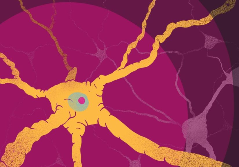
Gray matter contains many neuron cell bodies, dendrites, and synapsesstructures that receive, process, and integrate information. The cerebral cortex
(the brain’s outer layer) is gray matter, and it’s heavily involved in thinking, memory, attention, language, and planning. Deep gray matter structures
(like the thalamus and basal ganglia) help coordinate movement, attention, and the smooth flow of information.
If white matter is the delivery system, gray matter is the place where the packages get opened, interpreted, and turned into action. If MS damages both,
the brain can feel like it’s dealing with slow internet and a stressed-out CPU.
What MS is doing under the hood
Inflammation + immune misfires
MS is an immune-mediated disease of the central nervous system (brain, spinal cord, optic nerves). In MS, immune activity triggers inflammation that can
injure myelin, the cells that support myelin, and sometimes the axon itself. When this happens in a focal area, it can form a lesion (also called a plaque).
Demyelination is not the only story: axons and neurons can be injured too
MS has long been described as a demyelinating disease, but modern research and imaging show a broader picture: MS can involve neurodegenerationdamage to
axons and neuronsand brain volume loss (atrophy). Importantly, MS-related damage can affect both white matter and gray matter. That’s one reason MS is
often described as having both inflammatory and neurodegenerative components.
Translation: MS is not just “myelin getting peeled off.” It can be myelin injury plus downstream wear-and-tear on the nerve fibers and the cells doing
the thinking.
How MS affects white matter
White matter lesions: the classic MRI finding
White matter lesions are common in MS and often show up on MRI as bright areas on T2/FLAIR sequences. Lesions frequently appear in characteristic
locations such as near the ventricles (periventricular), near the outer edge of the brain (juxtacortical), in the brainstem/cerebellum, and in the
spinal cord. These patterns help clinicians distinguish MS from other causes of white matter changes.
Active vs older lesions (and why “new” matters)
Some lesions are “active,” meaning there’s ongoing inflammation. With contrast MRI (gadolinium), active lesions may enhance for a limited window of time.
Older lesions may not enhance, but they can still represent prior damage. Over time, some lesions can evolve in ways that suggest more permanent tissue injury.
Normal-appearing white matter isn’t always normal
Even when white matter looks okay on standard MRI, there can be subtle microscopic changesaltered myelin integrity, inflammation, or axonal injurythat
don’t show up as obvious lesions. Advanced MRI techniques (like diffusion imaging or magnetization transfer imaging) can detect changes that conventional
scans may miss.
What white matter damage can feel like
White matter disruption can affect how efficiently brain networks communicate. Depending on where lesions appear, people may experience:
- Vision problems (especially if the optic nerve pathway is involved)
- Numbness or tingling (sensory pathway disruption)
- Weakness or coordination problems (motor/cerebellar pathways)
- Slower processing speed (network efficiency takes a hit)
A key nuance: lesion count alone doesn’t perfectly predict symptoms. Some people have many lesions and function well; others have fewer lesions but more
disability. That’s where gray matter and brain atrophy enter the conversation.
How MS affects gray matter (the part people forget to invite to the conversation)
Cortical lesions: MS can affect the cortex
The cerebral cortex is gray matterand MS can cause demyelination there, too. Cortical lesions can be harder to detect on standard MRI, especially early
in the disease. Specialized imaging sequences and higher-field MRI can improve detection. Research has shown cortical demyelination can occur even in
early MS and may relate to clinical outcomes.
Deep gray matter: thalamus and basal ganglia involvement
Deep gray matter structures are heavily connected “hubs.” The thalamus, for example, helps route sensory and cognitive information. Atrophy in deep gray
matter has been associated with cognitive impairment and disability in many studies. When these hubs lose volume or integrity, symptoms can look like
slowed thinking, difficulty multitasking, fatigue that feels “brain-based,” and movement changes.
Gray matter atrophy: brain shrinkage beyond normal aging
Everyone loses some brain volume with age, but MS can accelerate brain volume loss. Gray matter atrophy (including cortical thinning and deep gray matter
shrinkage) is often more closely tied to disability progression than lesion counts alone. This is one reason clinicians and researchers increasingly pay
attention to “brain health” metrics over time, not just new lesions.
Why gray matter damage can track symptoms so well
Gray matter is directly involved in processing, memory, attention, mood regulation, and movement control. If MS affects gray matter, you may see:
- Cognitive changes (especially slowed processing speed, attention, working memory)
- Fatigue that feels more like “mental battery drain” than simple tiredness
- Mood changes (depression/anxiety can have multiple causes, but brain changes can contribute)
- Progressive disability even when new white matter lesions are less obvious
Think of it like this: white matter lesions can interrupt messages traveling across the brain, but gray matter damage affects the “processors” that turn
those messages into decisions and actions. When both are involved, symptoms can stack.
How white matter and gray matter damage are connected
Network effects and “downstream” degeneration
The brain runs on networks. If a white matter pathway is injured, the gray matter regions that depend on it can struggle. Over time, disrupted connections
may contribute to volume loss in connected gray matter regionsespecially in networks that are used constantly, like attention and motor circuits.
Inflammation can spill over into gray matter
Inflammation doesn’t always respect neat anatomical boundaries. Lesions near the cortex (juxtacortical lesions) may involve both white matter and adjacent
gray matter. Additional immune activity near the surface of the brain and in surrounding tissues has also been linked to cortical injury in MS.
What doctors look for: MRI clues in white and gray matter
Standard MRI: lesion activity and distribution
Clinicians use MRI to look for lesions in typical MS locations and to assess whether lesions are new or active. MRI is also used over time to see if
disease-modifying therapy (DMT) is reducing new inflammatory activity.
Advanced MRI: cortical lesions, microstructural damage, and atrophy
Many routine MRIs focus on lesion detection. But increasingly, clinicians and researchers use tools that estimate brain volume changes and highlight
subtle tissue injury. Some centers use sequences designed to better visualize cortical lesions, and some research settings use ultra-high-field MRI to
capture gray matter pathology more clearly.
Brain atrophy measurement: the slow-burn metric
Lesions can appear quickly, but atrophy is often a slower signal of cumulative injury. Tracking brain volume over time can provide insight into disease
progression and treatment effects, though it must be interpreted carefully (hydration, scanner differences, and timing can affect measurements).
Why disability can progress even when white matter lesions don’t “look worse”
This is a common and understandably frustrating experience: a person feels worsewalking is harder, thinking is slower, fatigue is heavieryet the MRI
report says, “No new lesions.” That can happen because:
- Gray matter injury may be progressing but is less visible on conventional MRI.
- Spinal cord damage can drive disability and may not be fully captured in a brain-only scan.
- Atrophy may reflect cumulative loss that doesn’t show up as “new spots.”
- Normal-appearing tissue may still have microstructural injury.
Some research in progressive MS also suggests that permanent disability can develop with relatively little classic cerebral white matter demyelination,
reinforcing that MS mechanisms can differ across people and across disease stages.
Can treatment protect white and gray matter?
DMTs: best at reducing inflammatory activity
Disease-modifying therapies are primarily designed to reduce relapses and new inflammatory lesions. Many studies also investigate whether DMTs slow brain
volume loss. While the details vary by medication and study design, the big picture is that preventing inflammatory injury early can help protect long-term
brain tissue.
Rehabilitation and brain-friendly habits: supporting the tissue you still have
MS care is bigger than medication. Strategies commonly used in comprehensive MS care include:
- Physical therapy for gait, balance, strength, and energy conservation
- Occupational therapy for daily tasks and cognitive strategies
- Cognitive rehabilitation (especially for processing speed and attention)
- Sleep optimization (sleep problems can amplify brain fog and fatigue)
- Managing vascular risks (blood pressure, diabetes, smoking) to support overall brain health
These don’t “cure” MS, but they can reduce symptom burden and help the brain run its remaining networks more efficientlylike upgrading the software when
the hardware is under stress.
Specific examples: how white and gray matter changes can show up day-to-day
Example 1: “I can walk fine, but my brain is buffering”
Someone may have relatively mild motor findings but noticeable cognitive slowingtaking longer to follow conversations, struggling to multitask, or feeling
overwhelmed in busy environments. This pattern can align with gray matter involvement, deep gray matter atrophy, or network inefficiency from widespread
white matter disruption.
Example 2: “My MRI is stable, but I’m slipping”
A person with progressive symptoms might not develop many new enhancing lesions, yet they may experience gradual worsening of mobility, endurance, or
manual dexterity. Atrophy and gray matter pathology can be part of the explanation, along with spinal cord involvement or long-term axonal injury.
Example 3: “My symptoms don’t match the number of lesions”
Lesion location matters more than raw count. A small lesion in a key pathway can cause big symptoms, while multiple lesions in less critical regions may
cause fewer noticeable problems. Gray matter injury and atrophy can further explain differences between scan appearance and real-life function.
When to contact a clinician
Contact your MS care team promptly if you notice new or worsening neurological symptomsespecially new vision loss, significant weakness, new balance
problems, or changes that interfere with daily function. Also ask about whether your imaging plan includes monitoring for brain volume changes, spinal cord
involvement, or cognitive screening if thinking or memory changes are affecting school, work, or home life.
Conclusion: the big takeaways
MS is often introduced as a white matter demyelinating diseaseand white matter lesions are a major part of it. But MS can also affect gray matter through
cortical lesions and deep gray matter injury, and those changes can be strongly tied to cognitive symptoms, fatigue, and disability progression.
The most useful way to think about MS-related brain change is “network health.” White matter damage can disrupt communication; gray matter damage can reduce
processing capacity; and atrophy can reflect cumulative injury over time. A good MS plan looks at all of it: relapse control, lesion monitoring, symptom
management, cognitive and physical function, and long-term brain health.
+500 WORDS: EXPERIENCES SECTION
Experiences: What living with white- and gray-matter changes can be like
When people talk about MS and the brain, the conversation often starts with MRI imagesbright spots, dark spots, “active” lesions, “stable” scans. But daily
life doesn’t happen inside an MRI machine. It happens while you’re trying to remember why you walked into the kitchen, follow a fast conversation, get through
a workday without feeling like your brain has run a marathon, or walk across a parking lot on legs that suddenly feel like they’re wearing invisible ankle weights.
One common experience tied to brain network changes is slowed processing speed. People describe it as “my thoughts are there, but they arrive late.”
In practical terms, it can look like needing extra time to answer questions, struggling to take notes while listening, or feeling overwhelmed when multiple people
talk at once. If gray matter hubs (like the thalamus) are under strainor if white matter pathways are slowing the flow of informationthe brain may still do the job,
just not at the same speed. That mismatch can be frustrating, especially because it’s often invisible to others. From the outside, you look fine. Inside, your brain
is asking everyone to please stop opening new tabs.
Another frequent experience is fatigue that feels neurological, not just “I didn’t sleep enough.” People sometimes say, “My body is tired, but my brain
is tired first.” This kind of fatigue may flare after mental effortlong meetings, studying, driving in heavy traffic, or even intense social time. It can feel like
your brain’s energy budget is smaller than it used to be, and once you overspend it, you pay interest for the rest of the day. When gray matter is involved, the
systems responsible for attention and effort can require more “fuel” to produce the same output. When white matter is involved, the brain may need more effort to
route signals around damaged pathways. Either way, the result can be the same: you’re exhaustedand it doesn’t always match how much you physically did.
People also describe changes in word-finding and working memory. You know what you want to say, but the word hides like it’s playing
a game of peekaboo. Or you can hold one piece of information in mind, but add a second task and the first one evaporates. These experiences can feel scary at first,
but many find that naming the pattern helps: “I’m not losing my intelligence; my brain is running on a slower, less consistent connection today.” For some, cognitive
screening and rehab strategies (like chunking information, using reminders, and reducing multitasking) make a noticeable difference in day-to-day confidence.
A final experience worth mentioning is the emotional side of “stable scans.” If you feel worse and the MRI report says “no new lesions,” it can feel invalidating.
But many people eventually learn a more accurate phrase: “No new lesions” doesn’t always mean “no new change.” Atrophy, spinal cord involvement, and gray matter
injury can progress in ways that are subtle on routine imaging. For many, the most helpful step is bringing concrete examples to appointmentswhat changed, when it
changed, and how it affects function. That information helps clinicians decide whether further imaging (including spinal cord MRI), cognitive testing, rehabilitation,
or treatment adjustments might be useful.
In short, living with MS-related white and gray matter changes can be a lesson in self-advocacy and strategy. Many people find relief in a combination of medical
care (to reduce inflammatory activity), rehabilitation (to strengthen function and build compensatory skills), and lifestyle supports (sleep, stress management,
movement within tolerance, and routines that reduce cognitive load). Your brain may not always feel predictable, but with the right supports, many people build a
life that’s not only manageablebut still meaningful, productive, and very much their own.