Table of Contents >> Show >> Hide
- Definition: What is the occipital lobe?
- Function: What does the occipital lobe do?
- How vision gets to the occipital lobe (quick pathway tour)
- Signs something’s wrong: Symptoms linked to occipital lobe problems
- Linked conditions: What can affect the occipital lobe?
- How clinicians evaluate occipital lobe problems
- Treatment and management (depends on the cause)
- When to seek urgent care
- Frequently asked questions
- Conclusion
- Real-life experiences and what people often notice (about )
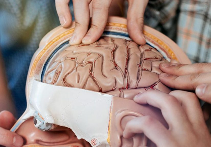
If your brain were a movie studio, the occipital lobe would be the editing bay, color-grading suite, and
“wait… was that a cat or a backpack?” department all rolled into one. It sits at the back of your head and is best known
for turning raw light signals into the visual world you experienceshapes, color, motion, depth, and (most days) the ability
to find your phone while it’s literally in your hand.
This article explains what the occipital lobe is, what it does, how it fits into the visual system, and which medical
conditions are commonly linked to itplus what symptoms should make you take things seriously (because “my vision is doing
something weird” is not a hobby).
Definition: What is the occipital lobe?
The occipital lobe is one of the four major lobes of the cerebral cortex. It’s located in the
posterior (back) part of the brain, roughly behind the parietal and temporal lobes. Its core job is
visual processingreceiving visual input and transforming it into meaningful perception.
Where it sits (and why that matters)
The occipital lobe is tucked beneath the back of the skull. Because it’s positioned where it is, it’s especially vulnerable
to some posterior circulation blood-flow problems (like certain strokes) and to head trauma
impacting the back of the head. Its location also helps explain why some occipital problems show up as “vision issues”
even when your eyes are perfectly healthy.
The visual cortex lives here
Inside the occipital lobe are specialized areas called the visual cortex. The most famous is the
primary visual cortex (often called V1 or Brodmann area 17), which sits
along the calcarine sulcus on the brain’s medial (inner) surface. Surrounding regionsoften discussed as
visual association areas (commonly tied to Brodmann areas 18 and 19)help interpret what V1 detects.
Blood supply: the posterior cerebral artery
The occipital lobe is mainly supplied by the posterior cerebral artery (PCA). This matters because
a PCA stroke can disrupt occipital function and cause classic visual symptoms like homonymous hemianopia
(loss of one side of the visual field in both eyes).
Function: What does the occipital lobe do?
The occipital lobe doesn’t just “see.” Your eyes detect light. Your occipital lobe turns that signal into information your
brain can use: edges, movement, patterns, and spatial layoutsthen hands it off to other brain networks to recognize objects,
read words, navigate a hallway, or admire a sunset without walking into a mailbox.
Primary visual processing (V1): building the basic picture
V1 receives visual input that has traveled from the retina to a relay station in the brain called the
lateral geniculate nucleus (LGN) in the thalamus. From there, signals reach the occipital lobe via
optic radiations. V1 helps map the visual world in an organized way (a “visual field map”), detecting
basics like contrast, orientation, and location.
Association visual processing: making vision useful
Once the basic signal is mapped, nearby visual areas help interpret it. Depending on the network involved, this can support:
- Color and form (telling a ripe strawberry from a decorative red ball)
- Motion detection (identifying that something is moving toward youimportant for sports and, you know, survival)
- Depth and spatial relationships (judging distance while driving or walking down stairs)
- Visual attention (deciding what to focus on when your environment is busy)
Working with other lobes: the “what” and “where/how” routes
Visual information doesn’t stay in the occipital lobe. It travels to other regions through two broad pathways:
- Ventral stream (“what” pathway): helps identify objects, faces, and complex patterns (more occipital-to-temporal).
- Dorsal stream (“where/how” pathway): supports spatial awareness, motion, and visually guided actions (more occipital-to-parietal).
That’s why occipital problems can sometimes look like “vision issues,” “reading issues,” or even “clumsiness” that’s actually
a visuospatial processing problem.
How vision gets to the occipital lobe (quick pathway tour)
Here’s the simplified route your visual signals follow:
- Retina detects light and converts it into neural signals.
- Optic nerve carries signals toward the brain.
- Optic chiasm partially crosses signals so each brain hemisphere receives information from both eyes.
- Optic tract carries signals to the thalamus.
- LGN (thalamus) relays and organizes input.
- Optic radiations project to the occipital lobe.
- Primary visual cortex (V1) begins cortical visual processing.
A key clinical detail: injuries behind the optic chiasm (in the tract, radiations, or occipital cortex) tend to cause
homonymous visual field defectsmeaning the same side of the visual field is affected in both eyes.
Signs something’s wrong: Symptoms linked to occipital lobe problems
Occipital issues often show up as changes in visual perception, not just “blurry vision.” Many people describe
it as “my eyes are fine, but my brain isn’t cooperating.”
Visual field loss (hemianopia and quadrantanopia)
If the occipital lobe is damagedespecially from a strokeone of the most classic symptoms is
contralateral homonymous hemianopia: losing the left half of the visual world (in both eyes) after a right-sided
brain injury, or vice versa. Smaller injuries can cause quadrantanopia (loss of a quarter of the visual field).
Quick guide: common patterns you might hear about
| Pattern | What it feels like | Often linked to |
|---|---|---|
| Homonymous hemianopia | Half the visual world is missing on the same side in both eyes | Occipital stroke, tumor, trauma (posterior to optic chiasm) |
| Quadrantanopia | A “slice” (quarter) of vision is missing | Damage along optic radiations or occipital cortex |
| Macular sparing | Central vision partly preserved even with field loss | Some occipital strokes (variation in blood supply/collaterals) |
Visual processing problems (when vision is there, but meaning is missing)
Some occipital and “posterior network” problems don’t erase the visual field but disrupt interpretation. People may:
- Struggle with reading even though their eyes can focus
- Have trouble judging distance (bumping into objects, misreaching for items)
- Feel overwhelmed in visually busy places (stores, crowds, patterned floors)
Cortical blindness
Cortical blindness can occur when both occipital cortices (or key visual pathways) are severely disrupted.
The eyes may be structurally normal, but the brain can’t process the signals. This is commonly associated with
posterior circulation strokes and other occipital injuries.
Visual hallucinations, flashes, and “special effects”
Occipital cortex irritation can produce positive visual phenomenaflashes, shapes, shimmering patternsor
negative phenomena like brief blindness in part of the field. Two common culprits:
-
Occipital seizures: often brief, may cause multicolored shapes, flashing lights, or transient field loss,
sometimes with eye movements or blinking. -
Migraine aura: often lasts longer (commonly minutes), may include zigzags, shimmering lines, or scotomas,
and can occur even without a headache in some people.
Linked conditions: What can affect the occipital lobe?
1) Posterior cerebral artery (PCA) stroke
A PCA stroke can injure the occipital lobe and is a classic cause of homonymous hemianopia. Some people notice
they keep bumping into doorframes on one side or “lose” words at the end of a line while reading. Because stroke is time-sensitive,
sudden new visual field loss should be treated as an emergencyespecially if it’s accompanied by weakness, confusion, severe headache,
trouble speaking, or imbalance.
2) Occipital lobe epilepsy
Occipital lobe seizures start in the visual processing region, so symptoms are often visual: flashing lights,
colored spots, field loss, or visual distortions. Some people also have rapid eye blinking or involuntary eye movements.
While occipital seizures are considered less common than temporal lobe seizures, they’re important because they can mimic
migraine aura or eye problems.
3) Migraine with aura (including aura without headache)
Migraine aura is thought to involve changes in brain activity that can affect the occipital cortex, producing visual symptoms.
Auras may include zigzag lines, shimmering patterns, blind spots, or temporary visual loss. Some people have aura without the
follow-up headache, which can be confusing (and is one reason new or unusual aura symptoms should be evaluated by a clinician).
4) Posterior cortical atrophy (PCA)
Posterior cortical atrophy is a rare, progressive neurodegenerative syndrome often associated with Alzheimer’s
pathology, where the back (posterior) parts of the brainincluding occipital and nearby regionslose function over time.
Early symptoms commonly involve visual processing: difficulty reading, judging distances, recognizing objects, or navigating.
5) Tumors and mass lesions
A tumor in or near the occipital lobe can press on visual cortex or pathways and cause gradual visual field defects, headaches,
seizures, or changes in visual perception. “Gradual and worsening” is a common clue here, though symptoms vary widely depending on
size and location.
6) Traumatic brain injury (TBI)
Falls, sports injuries, vehicle crashes, or blows to the back of the head can affect occipital tissue and trigger symptoms like
light sensitivity, trouble tracking movement, visual field changes, or visual fatigueespecially when combined with concussion-related
issues like headaches and concentration problems.
7) Inflammation, infection, or demyelination
Conditions that inflame or damage brain tissuelike encephalitis or certain demyelinating diseasescan also affect the occipital lobe,
sometimes producing visual symptoms, seizures, or broader neurologic changes depending on the pattern of involvement.
How clinicians evaluate occipital lobe problems
Because visual symptoms can come from the eye, optic nerve, or brain, evaluation often uses a “rule out the basics, then localize”
approach:
History and neurologic exam
- When did symptoms startsuddenly or gradually?
- Are symptoms constant, intermittent, or triggered (stress, sleep loss, flashing lights)?
- Any headache, weakness, speech changes, confusion, or seizure-like episodes?
Vision and visual field testing
Formal visual field testing (like perimetry) can map missing areas precisely. This is especially useful for
diagnosing hemianopia or quadrantanopia and tracking recovery over time.
Imaging and electrical testing
- CT or MRI can identify strokes, tumors, bleeding, inflammation, or structural injury.
- Vascular imaging (CTA/MRA) may be used if stroke is suspected.
- EEG may help if seizures are suspected.
Treatment and management (depends on the cause)
There isn’t one single “occipital lobe treatment.” Management is targeted to the underlying condition:
Stroke
Acute stroke treatment is time-critical and may involve clot-busting medications or procedures in eligible patients, plus
long-term risk reduction (blood pressure control, diabetes management, cholesterol management, smoking cessation, and appropriate
medications).
Seizures
Occipital epilepsy is typically managed with anti-seizure medications and lifestyle adjustments (sleep, stress management,
avoiding known triggers). Some cases need advanced evaluation if seizures are difficult to control.
Migraine with aura
Migraine treatment can include acute medications, preventive medications for frequent attacks, and trigger management.
New aura symptomsespecially if they’re sudden, prolonged, or very different from your usual patterndeserve medical review to
rule out more urgent causes.
Vision rehabilitation for field loss
Visual field loss from occipital injury can be life-changing, but rehabilitation can help. Strategies may include:
- Compensatory scanning training (learning systematic eye/head movements to cover the missing field)
- Occupational therapy for safe navigation and daily tasks
- Assistive tools (prism lenses in selected cases, environmental modifications)
When to seek urgent care
Seek emergency evaluation for sudden vision loss, new visual field loss, or sudden neurologic symptomsespecially if you have:
- Weakness or numbness on one side
- Difficulty speaking or understanding speech
- Severe, unusual headache
- Confusion, fainting, or seizure activity
Frequently asked questions
Can the occipital lobe affect reading?
Yes. Reading relies on visual processing networks, including posterior brain regions. Damage or degeneration in occipital or
posterior pathways can make reading slower, harder, or visually confusingeven if eyesight is otherwise “fine.”
Is occipital neuralgia the same thing as an occipital lobe problem?
Noeasy mix-up. Occipital neuralgia involves irritation of the occipital nerves in the scalp/neck region,
usually causing headache-like pain. The occipital lobe is brain tissue responsible for visual processing.
Similar name, completely different neighborhood.
Can vision recover after an occipital stroke?
Recovery varies. Some people improve naturally over time, and rehabilitation may help maximize function through compensatory strategies
and training. The best plan depends on the cause, severity, and overall neurologic health.
Conclusion
The occipital lobe is your brain’s visual enginetransforming raw signals into the meaningful world you navigate every day.
When it’s disrupted, symptoms often show up as visual field loss, visual distortions, trouble interpreting what you see, or
brain-based visual phenomena like migraine aura or occipital seizures. Because some causes (like stroke) are urgent, sudden new
visual changes should never be ignored. With accurate diagnosis and targeted treatmentplus vision rehabilitation when neededmany
people can adapt and improve function over time.
Real-life experiences and what people often notice (about )
Talking about the occipital lobe can sound abstract until you hear what occipital-related symptoms feel like in everyday life.
Below are common real-world experiences people reportshared here as practical examples, not as self-diagnosis tools.
1) “Half the world is missing… but I didn’t notice at first.”
One of the strangest things about a visual field cut (like homonymous hemianopia) is that your brain may try to “paper over”
the missing area. People often realize something is wrong indirectly: they keep clipping their shoulder on doorframes, they
leave food untouched on one side of a plate, or they can’t find a friend standing “right there” until they turn their head.
Reading can become unexpectedly exhausting because the eyes and head have to scan farther to capture the same line of text.
Many describe it as needing to “learn a new way of looking,” where deliberate scanning becomes a daily skilllike upgrading from
automatic to manual transmission.
2) “My vision turned into a screensaver.”
Migraine aura is often described with surprisingly consistent metaphors: shimmering zigzags, sparkles, a crescent-shaped blind
spot, or a moving distortion that slowly expands. People sometimes panic because it feels like an eye problemuntil they discover
it’s coming from brain-based visual processing. A common “aha” moment is realizing the effect appears in both eyes (even if it
feels stronger on one side), which can suggest a cortical origin rather than a single-eye issue. For some, aura happens without
head pain, which is equal parts relieving (“no headache!”) and confusing (“why am I seeing pixel fireworks?”).
3) “It was a flashthen it was gone.”
Occipital seizures can be brief and dramatic: sudden flashes, colored spots, or a rapid “light show” effect that lasts seconds
to minutes. Some people notice eye blinking or involuntary eye movements. Because these episodes can look like migraine aura,
the pattern matters: seizures are often shorter and may repeat in clusters. People frequently report that what scares them most
isn’t the visual symptom itselfit’s the unpredictability. That’s why accurate evaluation is so important: the right diagnosis
guides the right treatment and reduces unnecessary anxiety.
4) “My eyes test fine, but I can’t ‘make sense’ of what I’m seeing.”
Posterior processing issues (including posterior cortical atrophy) can feel like living in a world where the visual “data” arrives,
but the interpretation software is lagging. People may misjudge distances, struggle to find items in a cluttered drawer, get lost in
visually complex spaces, or find reading unusually difficult. A common frustration is being told, “But your vision is 20/20,” when
the problem is actually how the brain is processing visual information. When clinicians identify the pattern, it can be validating:
the person isn’t “careless” or “not trying”their brain is working harder to do the same tasks.
5) “Rehab feels awkward… until it doesn’t.”
Many people with occipital-related visual field loss describe rehabilitation as learning new habits: scanning left-to-right more
deliberately, pausing at intersections, arranging the home to reduce hazards, and using high-contrast cues. At first, these strategies
can feel clunkylike trying to dance while thinking about every step. Over time, repetition helps. The goal isn’t just “better test
results,” but safer walking, more confident reading, and fewer surprise collisions with the coffee table (which, to be fair, was
always a little too smug).
If any of these experiences sound familiarespecially if symptoms are new, sudden, or worseningthe safest move is to get a medical
evaluation. Brain-based vision issues are real, and they’re treatable or manageable in many cases when identified early.