Table of Contents >> Show >> Hide
Your kidneys are the behind-the-scenes coworkers who never take a lunch break: filtering waste, balancing fluids,
and keeping electrolytes from turning your bloodstream into a chemistry experiment. Most of the time, they do their job
quietlyuntil something feels “off” (swelling, high blood pressure, pain, abnormal labs, a transplant check, or a
suspicious ultrasound).
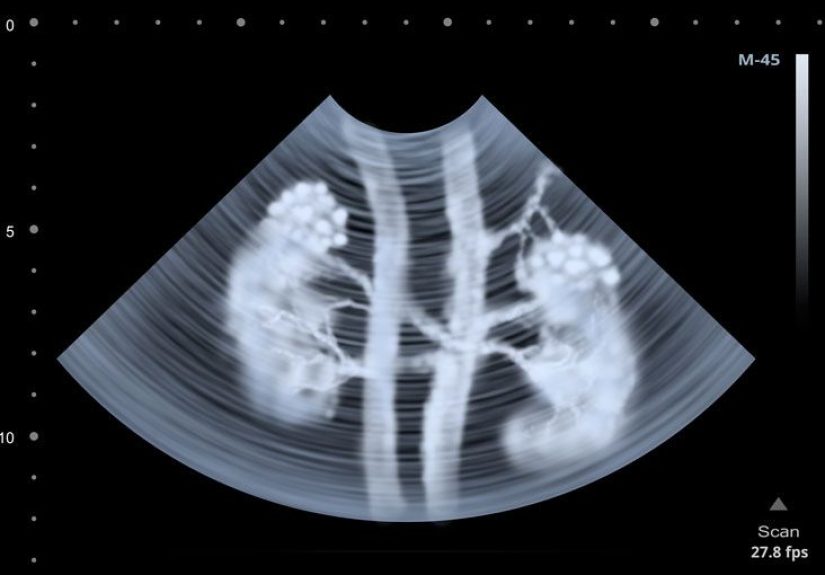
That’s where a renal scan (also called a kidney scan, renal scintigraphy,
or a renogram) can help. It’s a nuclear medicine imaging test that uses a tiny amount of
radiotracer and a special camera to show how well your kidneys are workingoften in real time.
Think of it as a “function movie,” not just a “structure snapshot.”
What a Renal Scan Is (and Isn’t)
A renal scan is an imaging exam that measures kidney function using a small amount of radioactive material
(a radioisotope or radiotracer) injected into a vein. A camera detects the tracer’s
gamma rays and builds images that show how blood flows into your kidneys, how the kidneys process the tracer, and how
urine drainage behaves.
What it’s not: a renal scan isn’t the same as a CT with contrast, an ultrasound, or an MRI. Those tests are
excellent at showing anatomy (stones, masses, cysts, hydronephrosis), but they may not answer the “How well is each kidney
actually working?” questionespecially when your provider needs split function or drainage dynamics.
Why Your Provider Might Order One
Providers usually order a renal scan when they want functional information that other tests can’t provide clearly.
Common reasons include:
- Suspected blockage or poor drainage (for example, hydronephrosis seen on ultrasound)
- Evaluating kidney blood flow or function patterns after surgery or injury
- Checking a transplanted kidney (perfusion and function monitoring)
- Investigating renovascular causes of high blood pressure (when kidney artery narrowing is suspected)
- Measuring how much each kidney contributes to overall function (differential/split renal function)
- Assessing kidney scarring after certain infections (commonly with cortical scans)
In plain English: if your care team needs to know whether a kidney is underperforming, obstructed, poorly perfused,
or contributing unevenlythis test is designed for that.
Types of Renal Scans
“Renal scan” is an umbrella term. Your specific test depends on the clinical question. Here are the most common categories:
| Type | What it’s best at | What to expect |
|---|---|---|
| Renal perfusion / functional imaging | Blood flow into the kidneys; overall function patterns | Images begin soon after injection; multiple images over ~20–30 minutes |
| Diuretic renal scintigraphy (often “Lasix renogram”) | Distinguishing true obstruction from slow-but-not-blocked drainage | Imaging before/after a diuretic; you may feel an urgent need to pee |
| ACE-inhibitor renal scintigraphy | Whether renal artery narrowing may be driving high blood pressure | Images before/after ACE-inhibitor medication |
| Renal cortical scintigraphy (commonly DMSA-based) | Cortical tissue function; scarring after infections | Injection first, then delayed imaging (often around ~2 hours later) |
| Dynamic renogram (general “renogram” approach) | Time-based uptake and washout; split function and drainage curves | Continuous/sequential images, usually 20–40 minutes for many protocols |
Your report might mention tracer names or protocol terms. Don’t panicthis doesn’t mean your kidneys have joined a sci-fi franchise.
It just reflects which radiotracer best answers the question (function, drainage, cortical detail, etc.).
How to Prepare
Preparation depends on the scan type, but these are common instructions:
- Hydration: You may be asked to drink extra fluids beforehand so the kidneys and bladder behave normally during imaging.
-
Medication review: Tell your care team about all medications, vitamins, and herbal supplements.
Some drugslike certain blood pressure medicines or NSAIDsmay affect results, and your provider may advise pausing them.
Don’t stop anything on your own. - Empty your bladder: Many protocols ask you to pee right before imaging.
- Pregnancy and breastfeeding: Always tell the team if you are pregnant, might be pregnant, or are breastfeeding, because nuclear medicine exams require extra planning.
- Comfort and metal: Wear comfortable clothing; remove jewelry or metal that could interfere with the camera setup.
Most adults don’t need fasting or sedation. Pediatric scans sometimes require special planning (and occasionally sedation), especially when staying still is a challenge.
Step-by-Step: What Happens During the Scan
Here’s a typical flow, with variations depending on the protocol:
1) Check-in and quick safety questions
You’ll confirm your medications, allergies, and pregnancy/breastfeeding status. If you’ve had a recent nuclear medicine study,
your provider may want to know because lingering tracer can affect accuracy.
2) IV placement and tracer injection
A technologist places an IV in your arm/hand, then injects the radiotracer. Most people feel only the needle stick and then…nothing dramatic.
The tracer itself usually doesn’t cause a “rush” sensation like some contrast agents can.
3) Imaging with a gamma camera
You’ll lie on (or sometimes sit at) an imaging table. The gamma camera positions near your abdomen/flank region.
Staying still mattersmotion can blur the functional “movie,” like filming a documentary during an earthquake.
Depending on the scan type, images may be taken:
- Immediately after injection to capture perfusion and uptake
- Over 20–40 minutes for many dynamic studies
- Later (delayed imaging) for cortical scans (often after a waiting period)
4) Optional medications during the scan (protocol-specific)
-
Diuretic (often furosemide/Lasix): Used to “push” urine flow and clarify whether drainage is truly blocked.
You may suddenly feel like your bladder has filed a strongly worded complaint. - ACE inhibitor: Used in specific hypertension workups to compare kidney function before and after medication.
5) Bathroom break, catheter, or special arrangements (sometimes)
Some protocols require a full or empty bladder at certain points. In certain pediatric or special cases, a catheter may be placed to keep the bladder empty
and improve accuracy, especially during diuretic studies.
6) After the scan
You can usually go home the same day. Many facilities recommend drinking fluids and urinating frequently for the next 24 hours to help flush the tracer.
Your IV site may be mildly soretreat it like any other small IV placement unless you notice worsening redness, swelling, or pain.
Time-wise, some renal scans take 30 minutes to 1 hour, while others may take longer depending on the protocol and whether delayed imaging is needed.
Understanding Results
Renal scan results can feel like reading a mix of medical jargon and a science fair poster. The good news: most reports revolve around a few core ideas.
1) Overall function and symmetry
Your provider looks at whether both kidneys take up and process tracer in a typical pattern. If one side is significantly slower or shows reduced uptake,
that can suggest decreased function, reduced blood flow, scarring, or obstructiondepending on the pattern.
2) Split (differential) renal function
Many renal scans estimate how much each kidney contributes to total function (often expressed as a percentage for left vs. right).
This is especially useful when planning surgery or evaluating a chronically obstructed system.
Example: if one kidney contributes far less than expected, your provider may focus on preserving the stronger side or addressing an obstruction earlier.
3) Drainage and “washout” (is it obstructed or just slow?)
In diuretic studies, the timing and slope of tracer washout help differentiate:
- Good drainage: tracer clears appropriately after the diuretic
- Delayed drainage without true obstruction: slow clearance, but improves with diuretic or positional changes
- Obstruction: persistent retention despite diuretic stimulation
A real-world scenario: an ultrasound shows hydronephrosis. That doesn’t automatically mean “blocked.”
A diuretic renogram helps answer whether urine is actually trapped (obstruction) or simply draining slowly without dangerous pressure buildup.
4) Perfusion (blood flow patterns)
Some protocols emphasize renal blood flow. Reduced perfusion can show up when the renal artery is narrowed or when there’s injury affecting the kidney’s blood supply.
In transplant monitoring, perfusion patterns can be particularly important.
5) Cortical tissue and scarring
Cortical scans are commonly used to evaluate renal cortical tissue and can help identify scarring patterns after infectionsespecially in pediatric care.
Delayed imaging improves the ability to “see” cortical detail.
How you’ll get the results
A nuclear medicine technologist and/or radiologist interprets the scan and sends a report to your ordering clinician. Your clinician then connects the imaging results
with your symptoms, labs, and other tests to decide what to do next.
Risks, Safety, and Special Situations
For most patients, a renal scan is considered low-risk. Still, it’s smart to know the key safety points:
-
Radiation exposure: There is a small amount of radiation from the tracer, and most of it leaves the body fairly quickly.
Your team uses the lowest dose that still answers the medical question. - Allergic reactions: Rare, but possible. Tell your team about any past reactions to medications or prior nuclear medicine studies.
- Pregnancy/breastfeeding: Extra precautions are needed. Your provider may recommend a different test or specific timing/temporary breastfeeding guidance.
- IV site issues: Mild soreness is common; worsening redness, swelling, or pain should be reported.
- Medications and accuracy: Some blood pressure meds, diuretics, NSAIDs, or recent nuclear medicine tracers can alter interpretation.
If you’re deciding between tests (ultrasound vs. CT vs. MRI vs. renal scan), remember: your provider is choosing based on the question.
“What does it look like?” and “How does it work?” are different problemsand sometimes you need both answers.
FAQ
Does a renal scan hurt?
The most uncomfortable part is usually the IV stick and holding still. If your study includes a diuretic, the urgency to urinate can be the main “event,”
but it’s temporary.
How long does it take?
Many scans take around 30–60 minutes, but some protocols take longer or include delayed imaging.
A common dynamic MAG3-style workflow may run closer to 1–2 hours from start to finish depending on the facility and protocol.
Will I be radioactive afterward?
You’ll have a small amount of tracer in your system temporarily. Most people are simply told to drink fluids and pee often to clear it.
If you’re pregnant or around pregnant people/infants, ask your facility for specific guidance.
Can I drive home?
Usually yesrenal scans are typically outpatient. If sedation is used (more common in certain pediatric settings), you’ll need a ride and follow facility instructions.
What should I do if the results are “abnormal”?
Don’t try to self-diagnose from the report wording alone. “Abnormal” can mean anything from mild drainage delay to reduced split function to perfusion issues.
Your clinician will interpret it in context and recommend next steps (repeat imaging, medication adjustments, urology/nephrology referral, or occasionally surgery planning).
Real-World Experiences (What People Commonly Notice)
Medical descriptions can be accurate and still feel emotionally unhelpfullike being told “a roller coaster is just physics.”
So here’s the human version of what many patients commonly experience (and what tends to surprise them) when they show up for a renal scan.
The waiting is often the longest part. Even when the scan itself is 30–40 minutes, check-in, IV placement, hydration instructions,
and any protocol timing can stretch the visit. For cortical scans (commonly used to look at scarring), some patients are surprised by a “pause” between injection
and imaging. That gap is normal: the tracer needs time to settle into the kidney tissue so the pictures can be meaningful.
The camera feels closer than expectedbut it’s not a tunnel. People who worry about claustrophobia often assume “scanner” means “MRI tube.”
A gamma camera is typically more open. It may hover near your abdomen/flank area, which can feel intimate in a “personal space bubble” way,
but it’s usually manageableand technologists can coach you through it.
The table can be cold and the stillness is real. A surprisingly common complaint is, “My back got stiff.”
You’re not being dramatic; holding still for imaging is uncomfortable for some people. A small tip that patients love:
wear comfy clothes, ask for a pillow or knee support if allowed, and do a quick bathroom break right before positioning if the protocol permits.
The diuretic step (if you have it) is the plot twist. If your scan includes a Lasix/diuretic portion, your bladder may suddenly feel like it’s
sending an urgent group text: “We need to talk. Now.” Some patients describe it as pressure rather than pain. It’s temporary, but it can feel intense,
especially if you’re instructed to remain still. Imaging teams usually anticipate this and will have a planbedpan, imaging toilet setup, or timing breaks,
depending on facility protocol.
Kids (and parents) often remember the IV more than the scan. In pediatric settings, families commonly say the hardest moment was the IV placement.
After that, distractions help: a stuffed animal, a favorite show, music, or a game. If a catheter is needed to keep the bladder empty, parents are often relieved to
learn it’s done for accuracynot because something “went wrong.” And yes, it can be emotionally tough in the moment, but it’s typically quick and carefully managed.
The “results anxiety” is real. Many people leave thinking, “If they took that many images, it must be bad.”
Not necessarily. Nuclear medicine studies generate lots of frames because they’re tracking function over timelike a flipbook of physiology.
It’s normal for interpretation to take a bit, because a radiologist is analyzing curves, symmetry, timing, and sometimes response to medications (diuretic or ACE inhibitor).
What patients often wish they’d known:
- Bring water (if allowed) and ask whether you should hydrate before arrival.
- Ask up front whether your scan includes a diuretic or delayed imaging so you can plan your time.
- If needles make you woozy, tell the staff earlythey have coping strategies.
- Afterward, fluids + frequent urination usually help you feel “back to normal” quickly.
Finally, a gentle reminder: this article can help you understand the test, but it can’t replace your clinician’s interpretation of your specific results.
The same scan pattern can mean different things depending on symptoms, labs, and other imagingso your next best step is always a results conversation with your care team.