Table of Contents >> Show >> Hide
- What Are Liver Lesions?
- Types of Liver Lesions
- 1) Benign (Noncancerous) Solid Lesions
- Hemangioma
- Focal Nodular Hyperplasia (FNH)
- Hepatic Adenoma (Hepatocellular Adenoma)
- Other Benign “Look-Alikes”
- 2) Benign Cystic Lesions
- Simple Liver Cyst
- Polycystic Liver Disease
- Hydatid (Echinococcal) Cyst
- 3) Infectious or Inflammatory Lesions
- Liver Abscess
- 4) Malignant (Cancerous) Lesions
- Hepatocellular Carcinoma (HCC)
- Cholangiocarcinoma
- Metastases (Cancer Spread to the Liver)
- Causes and Risk Factors
- Symptoms: When Do Liver Lesions Cause Trouble?
- How Liver Lesions Are Found and Diagnosed
- Treatment Options (By Lesion Type)
- Living With a Liver Lesion: Practical Tips That Actually Help
- When to Seek Urgent Care
- Real-World Experiences: What People Commonly Go Through (About )
- Conclusion
If you’ve ever opened an imaging report and seen the phrase “liver lesion,” you’ve probably felt your stomach drop.
Totally normal reaction. The word lesion sounds like something that should come with dramatic background music.
But here’s the twist: most liver lesions are benign (noncancerous), often found by accident, and many never cause a single problem.
In other words, lots of liver lesions are basically the “uninvited houseguest” who shows up, sits quietly in the corner,
and doesn’t eat your food.
This guide breaks down the most common types of liver lesions, what causes them, symptoms that actually matter,
how doctors diagnose them, and what treatment (if any) looks like. It’s educational, not a substitute for medical carebecause your liver
deserves personalized attention, not just internet vibes.
What Are Liver Lesions?
A liver lesion (also called a hepatic lesion or focal liver lesion) is an area of liver tissue
that looks different from the surrounding liver on imaging. “Lesion” is a broad label, not a diagnosis.
It can describe many thingslike a fluid-filled cyst, a benign tumor, an infection, scar-like changes, or (less commonly) cancer.
Liver lesions are frequently found during imaging for something unrelatedthink gallbladder pain, kidney stones, or a mystery stomach ache.
Modern ultrasound, CT scans, and MRI are very good at spotting tiny changes, which is helpful… and also the reason many people end up
Googling at 2 a.m.
Types of Liver Lesions
1) Benign (Noncancerous) Solid Lesions
Hemangioma
A hepatic hemangioma is the most common benign liver tumor. It’s essentially a cluster of blood vessels.
Most are small, discovered incidentally, and don’t need treatment. If they grow large, they can occasionally cause discomfort,
fullness, or nauseabut many people never notice them.
Typical plan: confirm it looks like a hemangioma on imaging, then watch only if your clinician thinks follow-up is needed.
(Translation: “We’re keeping an eye on it,” not “We’re panicking.”)
Focal Nodular Hyperplasia (FNH)
FNH is another common benign lesion, often considered a “regenerative” growthbasically liver tissue responding to blood-flow quirks.
It’s more common in women and often causes no symptoms. When imaging clearly identifies FNH, treatment usually isn’t needed.
You may hear “central scar” mentioned in imaging descriptions. That’s one of the clues radiologists look for,
especially on contrast MRI.
Hepatic Adenoma (Hepatocellular Adenoma)
A hepatic adenoma is benign, but it gets more attention than hemangioma or FNH because it has a higher chance of complications.
The big concerns are bleeding (especially if large) and a small risk of malignant transformation in certain subtypes.
Risk factors often include estrogen exposure (like oral contraceptives), anabolic steroid use, and metabolic factors such as obesity.
Because management depends on size, symptoms, and patient factors, adenomas are often monitored closely,
and some are treated with surgery or other procedures.
Other Benign “Look-Alikes”
Not every “lesion” is a true mass. Some are pseudolesions, such as focal fat deposition or focal fat sparing.
They can show up as odd-looking spots on imaging but aren’t tumors.
2) Benign Cystic Lesions
Simple Liver Cyst
A simple liver cyst is a fluid-filled sac and is usually harmless. Most cause no symptoms and require no treatment.
Larger cysts can sometimes cause bloating, pressure, or painmore “annoying roommate” than “dangerous intruder.”
Cysts with complex features (like internal septations, nodules, or unusual wall thickening) may need more imaging to rule out rare
precancerous or infectious causes.
Polycystic Liver Disease
Some people develop many cysts, sometimes linked with inherited conditions (including certain kidney disorders).
Treatment depends on symptoms and how cysts affect daily life. Many cases are managed conservatively;
intervention is usually reserved for significant symptoms.
Hydatid (Echinococcal) Cyst
This is caused by a parasite and is uncommon in the U.S., but it’s important because management is different and may involve medications
and/or procedures. Travel history and exposure risks matter here.
3) Infectious or Inflammatory Lesions
Liver Abscess
A liver abscess is a pocket of infection (pus) in the liver. This can cause fever, chills, right upper abdominal pain,
and feeling generally awful (technical term: “like garbage”). Abscesses require medical treatmenttypically antibiotics,
and sometimes drainage.
4) Malignant (Cancerous) Lesions
Hepatocellular Carcinoma (HCC)
HCC is the most common primary liver cancer. It usually arises in the setting of chronic liver disease, especially cirrhosis.
Risk is higher with chronic hepatitis B or C, long-term heavy alcohol use, and metabolic liver disease such as NAFLD/NASH.
In higher-risk patients, routine surveillance imaging is often recommended to catch HCC early.
Cholangiocarcinoma
This cancer begins in the bile ducts. Some forms occur inside the liver. Symptoms may include jaundice and itching,
though many cancers don’t cause symptoms until later stages.
Metastases (Cancer Spread to the Liver)
The liver is a common site for metastases from cancers elsewhere (such as colon, breast, lung, or pancreas),
largely because the liver filters a huge volume of blood. A personal history of cancer changes how clinicians interpret
a new liver lesion.
Causes and Risk Factors
Causes vary by lesion type, but these are common categories that influence risk and next steps:
- Underlying liver disease: cirrhosis, chronic hepatitis B or C, fatty liver disease (NAFLD/NASH), alcohol-related liver disease.
- Hormonal or medication exposure: estrogen-containing oral contraceptives, anabolic steroids, certain long-term hormone therapies.
- Metabolic factors: obesity, insulin resistance, high cholesterol, metabolic syndrome.
- Infection: bacterial spread from the abdomen, bloodstream infections, or (rarely) parasites.
- Cancer history: any prior malignancy raises suspicion for metastasis until proven otherwise.
- Inherited conditions: some genetic disorders are associated with adenomas or cystic disease.
Symptoms: When Do Liver Lesions Cause Trouble?
Most liver lesions cause no symptoms. The liver has a lot of “functional reserve,” meaning it can keep doing its job even when
something weird is happening in one small spot.
When symptoms do occur, they may include:
- Dull ache or discomfort in the right upper abdomen
- Feeling full quickly, bloating, or pressure (especially with larger cysts or masses)
- Nausea or decreased appetite
- Fever/chills (more suggestive of infection like an abscess)
- Unexplained weight loss, fatigue, or worsening appetite (can occur with cancer or advanced liver disease)
- Jaundice (yellowing of skin/eyes), dark urine, pale stools, itching (often bile-duct related or significant liver dysfunction)
Red Flags That Deserve Prompt Medical Attention
- Severe abdominal pain, dizziness, fainting (possible bleeding complicationespecially with certain adenomas)
- High fever with right-upper abdominal pain
- New or worsening jaundice
- Rapid belly swelling, confusion, or significant weakness
How Liver Lesions Are Found and Diagnosed
Diagnosis usually starts with imaging. The goal is to answer two big questions:
(1) What is it? and (2) Does it need treatment or monitoring?
Step 1: History and Risk Assessment
Clinicians don’t interpret a liver lesion in a vacuum. They consider liver disease risk factors (hepatitis, alcohol, fatty liver),
medication and hormone exposure, metabolic syndrome features, symptoms, and any history of cancer.
Blood tests may include liver enzymes and, in selected situations, tumor markers.
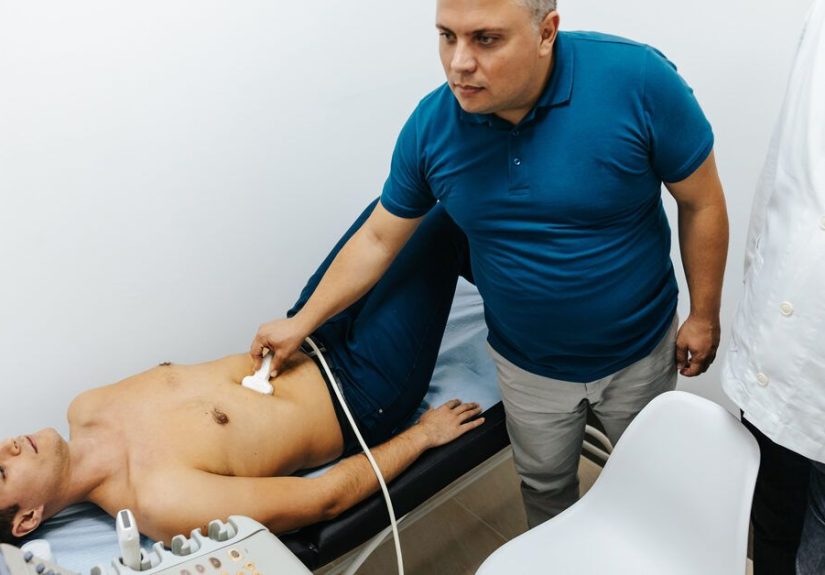
Step 2: Imaging (Ultrasound, CT, MRI, and Sometimes CEUS)
Many lesions are first seen on ultrasound. If the lesion isn’t clearly benign, the next step often involves
multiphase contrast-enhanced CT or contrast MRI. “Multiphase” is key: different lesions light up in different ways
depending on blood flow and how contrast moves through liver tissue.
MRI can be especially helpful for characterizing benign lesions and differentiating FNH from adenoma. In some cases,
hepatobiliary-specific contrast agents are used because they highlight lesions differently depending on bile duct features.
What About LI-RADS?
If you have cirrhosis or certain chronic liver conditions, imaging may use LI-RADS categories to estimate the likelihood of HCC.
Some lesions can be diagnosed on imaging alone in the right clinical context.
When Is a Biopsy Needed?
Biopsy isn’t always the first move because imaging can often identify classic benign patterns, and biopsies carry risks (like bleeding).
A biopsy may be considered if imaging is atypical, changing over time, or suspicious for malignancy.
Think of biopsy as the “we need a definitive answer” buttonnot the default setting.
Common Report Terms (So You Don’t Have to Speak Radiology)
- Hypodense/hypoattenuating: often means “less dense than surrounding tissue,” commonly cyst-like on CT.
- Hypervascular: a lesion that gets blood flow earlyseen in some benign lesions (FNH, adenoma) and some cancers.
- Incidental: found by accident during imaging for something else.
- Indeterminate: not clearly benign or malignant yetusually means “more imaging needed.”
Treatment Options (By Lesion Type)
Treatment depends on whether the lesion is benign vs malignant, whether it causes symptoms, its size and location,
and your overall liver health. Many people need no treatment at alljust confirmation and occasional monitoring.
Benign Lesions: Watchful Waiting, Sometimes Intervention
Hemangioma
Most hemangiomas require no treatment. If large or symptomatic, management may include monitoring and, rarely,
procedural or surgical options in select cases. In higher-risk patients (for example, those under surveillance for HCC),
clinicians may recommend closer imaging follow-up if a lesion is presumed to be a hemangioma.
FNH
If imaging confidently confirms FNH and you have no troubling symptoms, treatment is usually unnecessary.
Surgery is typically reserved for unusual cases: significant symptoms, uncertainty on diagnosis, or very large lesions causing compression.
Hepatic Adenoma
Adenomas are managed more actively because of bleeding risk and certain subtype risks. Common strategies include:
- Stopping estrogen exposure (for example, discontinuing estrogen-containing oral contraceptives when appropriate)
- Weight loss if overweight/obese, which may help reduce lesion growth in some cases
- Surveillance imaging to monitor size and features
- Resection or procedure if the lesion is large, symptomatic, growing, occurs in men, or shows higher-risk features
Your care team may also discuss subtyping and individualized riskbecause not all adenomas behave the same way.
Simple Liver Cysts
Most simple cysts need no treatmenteven if they’re largeunless they cause symptoms. If symptomatic, options may include
aspiration with sclerotherapy or surgical fenestration, depending on circumstances. Complex-appearing cysts often warrant
more detailed imaging first.
Liver Abscess
Abscesses require prompt medical care. Treatment commonly includes antibiotics and, when needed,
drainage guided by imaging. Most people improve significantly once the infection is addressed.
Malignant Lesions: A Different Playbook
For cancers (primary or metastatic), treatment depends on stage, liver function, and overall health. Options may include:
- Surgery (resection): removing the tumor when feasible
- Ablation: destroying tumors using heat or other methods
- Embolization: blocking blood flow to tumors (sometimes with chemotherapy or radioactive beads)
- Liver transplant: for selected patients with specific criteria, particularly for HCC
- Systemic therapy: targeted therapy, immunotherapy, or chemotherapy depending on cancer type
- Treatment of the primary cancer: in metastatic disease
Living With a Liver Lesion: Practical Tips That Actually Help
Whether your lesion is benign or still being evaluated, these steps can reduce stress and improve clarity:
- Ask for specifics: “What type of lesion do you suspect?” “What features make it look benign?” “What’s the follow-up plan?”
- Keep your reports and images: future comparisons are easier when nothing gets lost in the medical shuffle.
- Don’t DIY supplements: “liver cleanse” products can be surprisingly hard on the liver. Irony, thy name is detox tea.
- Support liver health: moderate or avoid alcohol, manage weight, control diabetes/lipids, and treat hepatitis if present.
- Know your risk level: surveillance recommendations differ a lot between low-risk and chronic liver disease patients.
When to Seek Urgent Care
Call a clinician urgently or seek emergency evaluation if you have severe abdominal pain, fainting, signs of significant bleeding,
high fever with abdominal pain, new jaundice, or confusionespecially if you have known liver disease.
Real-World Experiences: What People Commonly Go Through (About )
The most common “symptom” of an incidental liver lesion is not pain, nausea, or fatigueit’s anxiety.
Many people describe a very specific moment: you’re feeling fine, you get an ultrasound for something minor,
and suddenly a report says “lesion.” That single word can make it feel like your body has been quietly plotting behind your back.
A frequent experience is the confusing gap between discovery and clarity. You might hear:
“It’s probably a hemangioma,” followed by “We’ll confirm with an MRI.”
That in-between period can feel long, even if it’s only a couple of weeks.
People often say the hardest part wasn’t the lesionit was waiting while their brain auditioned for a role in a disaster movie.
Then comes the alphabet soup of imaging: “arterial phase,” “portal venous phase,” “delayed phase,” sometimes even “hepatobiliary contrast.”
Patients often report that once a clinician explains the basicshow benign lesions tend to have recognizable enhancement patterns
their stress drops quickly. Understanding that “more imaging” usually means “better identification,” not “we found something terrible,”
can be a game-changer.
People with benign findings often describe relief mixed with a strange sense of anticlimax:
“So… we’re just leaving it there?” Yes, sometimes the best treatment is literally doing nothing.
Many patients say it helped to reframe the lesion as a scar-like quirk or a harmless birthmark of the liver.
Not cute, exactlybut harmless.
Experiences differ when the lesion is an adenoma or a symptomatic cyst. Some patients describe a practical checklist:
stop estrogen-containing contraceptives (with guidance), adjust medications, work on weight and metabolic health,
and schedule follow-up imaging. For many, it feels empowering to have concrete steps.
Others find it frustratingespecially if the lesion was found when they felt perfectly healthy.
It can be emotionally weird to “treat” something that never bothered you until a scan pointed at it.
A smaller group experiences physical symptoms: fullness after meals, a persistent ache, or pressure that’s hard to describe.
Those with large cysts sometimes say their symptoms were dismissed as “just digestion” until imaging showed a cause.
After treatment (like drainage or fenestration), many report a noticeable improvement in comfort and appetite,
plus a surprising bonus: breathing and bending over can feel easier when a large cyst is no longer pushing on nearby structures.
For people with chronic liver disease, the experience can be more intense because surveillance is routine and the stakes can be higher.
Many describe becoming “fluent” in their own health datatracking ultrasound dates, lab trends, and follow-up plans.
In that context, a new lesion can feel scary, but structured surveillance can also provide reassurance:
problems are more likely to be caught earlier, when more options exist.
Across all these stories, the most helpful theme is consistent: the best next step is almost always
clarificationbetter imaging, a specialist opinion when needed, and a plan you understand.
Liver lesions are a category, not a verdict. And your goal isn’t to become a radiologist overnightjust to get the right information
and the right follow-up.
Conclusion
“Liver lesion” is an umbrella term covering many possibilitiesfrom simple cysts and hemangiomas to infections and cancer.
The good news is that most incidental liver lesions are benign and don’t require treatment.
The important part is proper characterization with appropriate imaging and risk-based follow-up.
If you’ve been told you have a liver lesion, focus on the practical questions: What type is it likely to be?
Do you have risk factors for liver disease or cancer? What imaging (if any) is needed next? And what symptoms should you watch for?
With clear answers, this often becomes a manageable, boring medical footnotewhich is exactly what you want.