Table of Contents >> Show >> Hide
- What Ocular Hypertension Is (and Isn’t)
- Why Eye Pressure Goes Up
- Risk Factors: Who’s More Likely to Have It (or Progress to Glaucoma)
- Symptoms: Why It’s Usually a “Quiet” Condition
- How Ocular Hypertension Is Diagnosed
- Treatment: When to Treat and What It Looks Like
- Everyday Habits That Support Eye Health (Without “Miracle Cure” Energy)
- Frequently Asked Questions
- Conclusion: The Big Picture
- Experiences: What Living With Ocular Hypertension Can Feel Like (and Why That Matters)
If you’ve ever been told you have “high eye pressure,” you might’ve pictured your eyeballs wearing tiny weightlifting belts.
The good news: ocular hypertension usually doesn’t come with dramatic symptoms or instant vision loss.
The not-so-fun news: it can raise your risk of glaucoma, which can damage the optic nerve over time.
This guide breaks down what ocular hypertension is, why it happens, how doctors diagnose it, and what treatment looks likewithout the doom-and-gloom vibe.
(Think: helpful, not horrifying.) And yes, we’ll talk about what living with it can actually feel like, because “asymptomatic” doesn’t mean “emotionless.”
What Ocular Hypertension Is (and Isn’t)
High eye pressure vs. glaucoma
Ocular hypertension means your intraocular pressure (IOP)the fluid pressure inside your eyeis higher than what’s typically considered normal,
but there’s no clear evidence of optic nerve damage and no vision loss on testing.
In other words, your pressure is elevated, but the “wiring” (the optic nerve) hasn’t shown damage.
Glaucoma, on the other hand, is a disease where the optic nerve gets damagedoften associated with elevated IOP, but not always.
Some people develop glaucoma with “normal” pressures (normal-tension glaucoma), and some people live with higher pressures and never develop glaucoma.
That’s why ocular hypertension is often described as being a glaucoma risk factor and a reason for careful monitoringnot a diagnosis of glaucoma by itself.
What counts as “high” eye pressure?
Eye pressure is measured in millimeters of mercury (mmHg). Many references place “typical” IOP somewhere around 10–21 mmHg.
Ocular hypertension is often defined as IOP readings consistently above that range, usually > 21 mmHg, without optic nerve damage.
But numbers aren’t the whole story: corneal thickness, measurement method, time of day, and your personal risk factors all matter.
Why Eye Pressure Goes Up
A quick tour of your eye’s “plumbing”
Your eye constantly makes a clear fluid called aqueous humor.
That fluid nourishes tissues in the front of your eye and normally drains out through a tiny “drainage angle,”
mainly via the trabecular meshwork.
If fluid doesn’t drain efficientlyor if it’s produced faster than it can exitpressure can rise.
Common causes and contributors
Ocular hypertension can be primary (no single identifiable cause) or secondary (linked to another condition or trigger).
Here are some of the usual suspects:
-
Reduced drainage (open-angle issues):
The angle may be “open,” but the trabecular meshwork doesn’t let fluid out as efficiently as it should.
This is common and often has no obvious day-to-day symptoms. -
Steroid response:
Corticosteroids (eye drops, injections, creams around the eyes, inhalers, or systemic steroids) can raise IOP in some people.
The longer or stronger the steroid exposure, the more likely pressure can climbespecially in “steroid responders.” -
Pigment dispersion syndrome:
Pigment granules from the iris can shed and clog the drainage system, nudging pressure upward. -
Pseudoexfoliation syndrome:
Flaky protein-like material can build up in the front of the eye and interfere with outflow, increasing IOP and glaucoma risk. -
Eye inflammation (uveitis) or injury:
Inflammation, trauma, or bleeding inside the eye can affect outflow and raise pressure. -
Post-surgical changes:
Some eye surgeries can alter drainage or inflammation patterns, influencing IOP.
Risk Factors: Who’s More Likely to Have It (or Progress to Glaucoma)
Two people can have the same IOP reading and very different risk profiles.
Eye doctors look at a cluster of risk factors to estimate your chance of developing primary open-angle glaucoma over time.
Risk factors often linked to higher glaucoma risk in ocular hypertension
- Higher baseline IOP (not just one readingpatterns matter)
- Older age
- Thinner central corneal thickness (CCT) (affects both measurement and risk prediction)
- Larger optic nerve cup-to-disc ratio or suspicious optic nerve appearance
- Family history of glaucoma
- Race/ethnicity (for example, Black/African American individuals have higher risk for primary open-angle glaucoma)
- Myopia (nearsightedness) in some cases
- Coexisting eye conditions like pigment dispersion or pseudoexfoliation
A landmark clinical trial (the Ocular Hypertension Treatment Study) helped clinicians quantify risk and showed that lowering IOP can reduce the chance of developing glaucoma in certain patients.
The key takeaway wasn’t “treat everyone”it was “treat the people who are most likely to benefit.”
Symptoms: Why It’s Usually a “Quiet” Condition
Here’s the tricky part: ocular hypertension usually has no symptoms.
No warning sirens. No blinking neon signs. Often, the only way to find it is during a routine eye exam.
If symptoms do happen, they’re usually related to something else going on (like inflammation, trauma, or a sudden change in eye anatomy).
Seek urgent eye care if you have severe eye pain, sudden blurred vision, halos around lights,
headache with nausea, or a rapid drop in visionthese can be signs of an eye emergency that needs prompt evaluation.
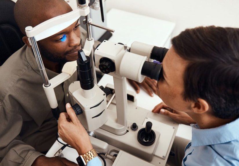
How Ocular Hypertension Is Diagnosed
Diagnosing ocular hypertension is less like a single “ta-da!” moment and more like assembling a puzzle.
Your eye doctor will usually confirm elevated pressure and check whether any glaucoma-related damage is present.
Tests you may see during an evaluation
- Tonometry: Measures IOP. Goldmann applanation tonometry is often considered the reference standard, but other devices exist (including “air puff” methods).
- Pachymetry: Measures corneal thickness. A thicker cornea can make IOP readings look higher than they truly are; a thinner cornea can mask higher pressure.
- Gonioscopy: Lets the doctor look at the drainage angle to confirm whether it’s open and assess for angle abnormalities.
- Dilated optic nerve exam: The doctor examines the optic nerve for suspicious changes.
- Optic nerve imaging (often OCT): Helps track subtle structural changes over time.
- Visual field testing: Checks for functional vision loss patterns typical of glaucomaoften before you notice changes in daily life.
Many clinicians also repeat measurements at different visits (and sometimes different times of day), because IOP can fluctuate.
If you’re thinking, “So… I need more than one data point?”yes. Your eyes are not a one-snapshot kind of situation.
Treatment: When to Treat and What It Looks Like
Treatment is usually about reducing the risk of glaucoma, not “curing” ocular hypertension in a permanent way.
Some people are managed with observation only; others benefit from pressure-lowering therapy.
The decision depends on your overall risk profile, your optic nerve appearance, test results, and how high your IOP runs over time.
Option 1: Monitoring (a.k.a. “watchful waiting,” but with real structure)
If your risk is lowhealthy-looking optic nerves, normal visual fields, and modest pressure elevationyour doctor may recommend monitoring without treatment.
That doesn’t mean “ignore it.” It means:
- Regular eye exams on a schedule tailored to your risk
- Periodic optic nerve imaging and visual field tests
- Tracking IOP trends (not just one-off numbers)
Option 2: Prescription eye drops
Eye drops are the most common first step when treatment is recommended.
Different medications lower IOP by either helping fluid drain out or reducing fluid production.
Common classes include:
-
Prostaglandin analogs (often once daily):
Increase outflow. Examples include latanoprost.
Possible side effects: redness, irritation, eyelash growth, and gradual iris or eyelid darkening in some people. - Beta-blockers: Reduce fluid production. Not ideal for everyone (for example, certain heart or lung conditions may matter).
- Alpha agonists: Reduce production and may increase outflow.
- Carbonic anhydrase inhibitors: Reduce production (available as drops and, less commonly, oral meds in select situations).
- Combination drops: Two medications in one bottlefewer bottles, more convenience, sometimes better adherence.
A practical tip: if you’re on drops, technique matters.
Many doctors recommend closing the eye gently and pressing the inner corner of the eyelid (near the nose) for about a minute after instilling drops.
This may help reduce how much medication drains into the tear duct and bloodstream.
Ask your eye doctor to demonstrate; it’s one of those “small change, big payoff” things.
Option 3: Laser trabeculoplasty (often SLT)
Selective laser trabeculoplasty (SLT) is a laser procedure that targets the drainage tissue to improve outflow and lower IOP.
It’s used for open-angle glaucoma and can also be used for ocular hypertension in appropriate patients.
Why some people like SLT:
- No daily drop routine (at least for a while)
- Can lower IOP meaningfully in many patients
- Often performed in an outpatient setting
Tradeoffs:
- Effect can wear off over time (and may need repeat treatment or drops later)
- It’s not a fit for every eye anatomy or glaucoma type
Option 4: Surgery (less common for ocular hypertension alone)
Surgery is usually reserved for cases where pressure is very hard to control or if glaucoma develops and progresses despite other treatments.
Minimally invasive glaucoma surgeries (MIGS) may be considered in certain scenarios, often alongside cataract surgery, but management is highly individualized.
Everyday Habits That Support Eye Health (Without “Miracle Cure” Energy)
There’s no proven diet or supplement that replaces medical monitoring or treatment for ocular hypertension.
But there are sensible habits that support overall eye health and help you avoid preventable pressure spikes or missed diagnoses:
- Keep your follow-up appointments (boring, yes; protective, also yes).
-
Tell your doctor about steroid useincluding inhalers, skin creams near the eyes, and “leftover” prescription drops.
Steroids are useful medications, but they’re not a DIY hobby. - Move your body: regular exercise supports cardiovascular health and may help overall wellness.
- Know your family history and share itglaucoma risk can run in families.
- Ask about caffeine and lifestyle triggers if your IOP fluctuatessome people notice patterns, and your clinician can guide what matters medically.
Frequently Asked Questions
Can ocular hypertension go away?
Sometimes elevated pressure is temporaryespecially if it’s caused by something reversible like steroid use or inflammation.
But many cases are chronic and need long-term monitoring.
The goal is to keep pressure at a level that protects your optic nerve over your lifetime.
If I have ocular hypertension, will I definitely get glaucoma?
No. Many people with ocular hypertension never develop glaucoma.
Risk depends on factors like baseline IOP, corneal thickness, optic nerve appearance, age, and family history.
That’s why individualized risk assessment is so important.
What’s the “target pressure”?
Target pressure isn’t one universal numberit’s a personalized goal set by your eye doctor, based on your risk and how your optic nerve looks over time.
Some treatment approaches aim for a percentage reduction in IOP (often around 20% in major studies), plus a pressure range that seems safest for your particular eyes.
Do eye drops have long-term side effects?
Most people tolerate drops well, but side effects can happen and depend on the medication.
Some are local (redness, dryness, stinging), and some can be systemic (especially with certain drop classes).
If a drop bothers you, don’t just quittell your doctor.
There are often alternatives.
How often do I need checkups?
It varies. Low-risk patients may be monitored less frequently than higher-risk patients.
Your clinician will base follow-up timing on your IOP level, test results, optic nerve status, and overall risk profile.
Conclusion: The Big Picture
Ocular hypertension is a “numbers-up, damage-not-yet” situation.
The pressure inside the eye is higher than expected, but the optic nerve still looks healthy and visual field testing hasn’t shown glaucoma-related loss.
Because ocular hypertension can increase the risk of developing glaucoma, the smartest approach is a personalized plan:
confirm the diagnosis with proper testing, assess risk factors, and decide whether monitoring, drops, laser treatment, or another strategy makes the most sense.
If there’s one takeaway worth taping to your bathroom mirror (right next to “floss,” which we both know you ignore sometimes):
ocular hypertension is manageableespecially when it’s found early and followed consistently.
Regular eye care can help protect your vision long-term.
Medical note: This article is for general educational purposes and doesn’t replace personalized medical advice.
If you’ve been told you have elevated eye pressureor you have new eye symptomsschedule a visit with an optometrist or ophthalmologist.
Experiences: What Living With Ocular Hypertension Can Feel Like (and Why That Matters)
Ocular hypertension has a weird social life: it’s often invisible, but it can still take up a lot of mental space.
Many people first learn they have it during a totally routine appointmentmaybe you went in for new glasses, or you finally replaced those scratched-up lenses
you’ve been “meaning to swap” since the last presidential administration.
Then your eye doctor casually says, “Your eye pressure is a bit high,” and suddenly you’re thinking,
“Wait… do my eyes have blood pressure now?”
One common experience is the anxiety of having a condition that doesn’t “feel” like anything.
If your knee hurts, you can point to the knee. If you have a fever, you can measure it.
With ocular hypertension, you’re often asked to care about a number you can’t sense.
That can make it tempting to treat follow-up visits like optional homework.
But many people say that once they understand the goalprotecting the optic nerve over decadesthe appointments feel less like busywork and more like
long-term vision insurance.
Another real-life theme: IOP numbers can fluctuate, and that can mess with your confidence.
Maybe your first reading was high, the second was lower, and now you’re wondering whether the first was “wrong.”
In reality, pressure can vary based on time of day, measurement method, stress, and simple biological variability.
Some people find it reassuring when their doctor explains the plan in trends: “We’re watching your pattern, not panicking over one number.”
That approach helps turn a scary reading into a manageable storyline.
For people who start drops, the learning curve is real.
It’s not just “put liquid in eye.” It’s remembering the schedule, using the right technique, keeping bottles clean, and figuring out where drops live
(bathroom? bedside table? backpack?) so you don’t miss doses.
Some folks describe the first week as surprisingly emotional: you may feel older than you are, or annoyed that your eyes now require a daily routine.
Others feel relievedlike they finally have a concrete action step instead of a “wait and see” plan.
Side effects can also shape the experience.
A little redness or stinging can feel like a big deal when you weren’t expecting it.
Some people worry the redness looks like they’re exhausted, sick, or crying (when they’re just… medicated).
The good news is that doctors can often switch medications, adjust timing, or consider laser options if drops are uncomfortable.
People who do best long-term often share a similar strategy: they report side effects early instead of silently suffering or quitting.
Then there’s the “label” effect.
Being called a “glaucoma suspect” can sound like you’re wanted in a medical crime drama.
Many patients say it helps when clinicians explain what the label actually means: it’s a risk category, not a verdict.
And that framing can be empoweringbecause risk is something you can manage with monitoring and treatment.
Finally, a lot of people find comfort in building a simple routine:
keep appointments, track drops, ask what your corneal thickness means for your readings, and understand your personalized risk.
It’s not glamorous, but it’s effective.
Ocular hypertension is often a long gameand people who treat it like a long game usually feel more in control, less blindsided, and more confident
that they’re doing the right things to protect their vision.