Table of Contents >> Show >> Hide
- What Is a Pancreatic Pseudocyst?
- What Causes a Pancreatic Pseudocyst?
- Risk Factors That Raise the Odds
- Pancreatic Pseudocyst Symptoms
- How Doctors Diagnose a Pancreatic Pseudocyst
- Pseudocyst vs. Other Pancreatic Cysts
- Why Timing Matters in Diagnosis
- When to Seek Medical Care Right Away
- What the Diagnosis Often Means for Patients
- Experience Section: What Living Through This Can Feel Like
- Conclusion
The term pancreatic pseudocyst sounds like something a villain would yell in a hospital drama, but the condition itself is very real. A pancreatic pseudocyst is a pocket of fluid that forms in or around the pancreas, usually after the pancreas has been irritated, inflamed, or injured. Most often, that injury comes from pancreatitis. In plain English: the pancreas gets angry, digestive fluids leak where they do not belong, and the body walls off the mess.
That wall is what makes a pseudocyst different from a true cyst. A true cyst has a lining made of epithelial cells. A pseudocyst does not. Instead, it is surrounded by fibrous or inflammatory tissue, more like the body’s improvised repair job than a neatly constructed storage unit. Not elegant, but very on-brand for human anatomy.
This matters because pancreatic pseudocysts are usually benign and often tied to a recent episode of pancreatitis, while other pancreatic cysts may have very different causes and, in some cases, cancer potential. So when a doctor sees “fluid collection near the pancreas,” the next step is not to panic. The next step is to figure out exactly what kind of lesion it is, why it formed, and whether it is causing trouble.
What Is a Pancreatic Pseudocyst?
A pancreatic pseudocyst is an encapsulated collection of pancreatic fluid, enzymes, inflammatory material, and sometimes a small amount of blood or tissue debris. It usually develops after acute pancreatitis, chronic pancreatitis, or pancreatic trauma. In many cases, it does not show up immediately. Instead, it tends to mature over several weeks, often around four weeks after the original attack of pancreatitis.
That timing is important. Early after acute pancreatitis, doctors may see a fluid collection that has not yet developed a mature wall. Once a defined capsule forms and the collection contains mostly fluid rather than solid necrotic material, it is more consistent with a pseudocyst. This is one reason imaging and timing go hand in hand. A scan taken too early may tell one story; a later scan may tell a much clearer one.
Pancreatic pseudocysts can vary in size. Some are small and barely noticeable. Others grow large enough to press on nearby organs, ducts, or blood vessels. When that happens, symptoms tend to get a lot more dramatic, and a patient who was merely uncomfortable may suddenly feel miserable.
What Causes a Pancreatic Pseudocyst?
1. Acute pancreatitis
The most common cause of a pancreatic pseudocyst is acute pancreatitis. Acute pancreatitis is a sudden inflammation of the pancreas that can happen when digestive enzymes activate too early and begin damaging pancreatic tissue. Two major causes of acute pancreatitis in the United States are gallstones and heavy alcohol use. Gallstones can block the pancreatic or bile ducts. Alcohol can directly injure pancreatic tissue and promote recurrent inflammation. Either way, the pancreas loses its cool, and fluid may leak into the surrounding area.
2. Chronic pancreatitis
Chronic pancreatitis is long-term inflammation that causes progressive damage to the pancreas. In this setting, scarring and duct obstruction can lead to repeated leaks of pancreatic fluid. Pseudocysts are especially common in people with chronic pancreatitis because the injury is not a one-time event. It is a repeat offender.
3. Pancreatic trauma
Direct injury to the pancreas can also lead to a pseudocyst. This may happen after blunt abdominal trauma, certain procedures, or surgery involving the pancreas. Trauma can disrupt the pancreatic duct, and once enzyme-rich fluid escapes, nearby tissues become inflamed. The body then tries to contain the leak by building a fibrous wall around it.
4. Duct disruption and leakage
Underneath all of these causes is one main mechanical problem: pancreatic fluid escapes from the ductal system. That fluid is packed with digestive enzymes. These enzymes are useful when they are in the small intestine helping digest lunch. They are much less charming when they spill into surrounding tissues and begin irritating everything in sight.
Risk Factors That Raise the Odds
Because pseudocysts are usually a complication of pancreatitis, the major risk factors are often the same ones that increase the risk of pancreatitis in the first place. These can include gallstones, heavy alcohol use, recurrent pancreatitis, chronic pancreatitis, high triglyceride levels, abdominal trauma, certain medications, and structural problems affecting the pancreatic or bile ducts.
That does not mean everyone with pancreatitis will develop a pseudocyst. Many people do not. But the risk rises when pancreatitis is severe, recurrent, or complicated by duct injury.
Pancreatic Pseudocyst Symptoms
Some pancreatic pseudocysts cause no symptoms at all. They are found incidentally on imaging ordered for pancreatitis follow-up or abdominal pain. In those cases, the patient may be surprised to learn a pseudocyst is even there. The pancreas, apparently, loves surprise plot twists.
When symptoms do appear, they usually reflect one of three things: ongoing inflammation, pressure on nearby structures, or a complication such as infection or bleeding.
Common symptoms
The most common symptom is upper abdominal pain, often centered in the epigastric area and sometimes radiating to the back. The pain may be persistent, dull, or pressure-like, though it can also be sharp. Nausea and vomiting are also common, particularly if the pseudocyst is associated with recent pancreatitis or is large enough to compress part of the stomach or small intestine.
Other symptoms may include abdominal bloating, a feeling of fullness, early satiety, poor appetite, and unintentional weight loss. A large pseudocyst can sometimes be felt as a mass in the upper abdomen, though that is less common.
Symptoms caused by pressure on nearby organs
If the pseudocyst presses on the stomach or duodenum, a person may feel full quickly after eating, vomit, or have trouble tolerating food. If it compresses the bile duct, jaundice may develop, causing yellowing of the skin or eyes, dark urine, pale stools, or itching. Compression of blood vessels is less common but more serious, particularly if bleeding develops.
Symptoms that suggest a complication
Fever, chills, worsening abdominal pain, and increasing illness can suggest infection. Sudden severe pain, dizziness, fainting, or signs of internal bleeding can point to hemorrhage or rupture, which are medical emergencies. Some pseudocysts also become infected and turn into or coexist with an abscess-like process. That is when the situation moves from “needs evaluation” to “stop reading and call a doctor.”
How Doctors Diagnose a Pancreatic Pseudocyst
Diagnosing a pancreatic pseudocyst is not just about spotting a fluid pocket on a scan. Doctors need to match imaging findings with symptoms, medical history, lab results, and timing. A round blob of fluid near the pancreas is not automatically a pseudocyst. It has to make sense in context.
1. Medical history and physical exam
Doctors usually start by asking whether the patient recently had acute pancreatitis, has chronic pancreatitis, uses alcohol heavily, had gallstones, or suffered abdominal trauma. The timeline matters. A new fluid collection seen days after acute pancreatitis may not yet qualify as a mature pseudocyst. A collection seen weeks later is more convincing.
During the physical exam, clinicians look for upper abdominal tenderness, distension, fever, jaundice, or signs of dehydration or systemic illness. If symptoms are severe, they also assess for possible complications such as infection or bleeding.
2. Blood tests
Blood work does not diagnose a pancreatic pseudocyst by itself, but it helps build the picture. Doctors may order amylase and lipase levels, especially if pancreatitis is still active or suspected. They may also check electrolytes, glucose, liver tests, triglycerides, kidney function, and a complete blood count.
If the white blood cell count is high or inflammatory markers are elevated, infection becomes more concerning. If bilirubin and alkaline phosphatase are elevated, that may suggest bile duct compression or another biliary problem. So while blood tests rarely deliver the final answer alone, they often tell doctors how worried they should be and what else may be going on.
3. Imaging tests
CT scan
Contrast-enhanced CT is one of the most commonly used tests for pancreatic pseudocysts. It can show the size, location, wall, and relationship to nearby organs. CT is especially helpful when a patient has ongoing pain after pancreatitis or when doctors need to assess complications.
Ultrasound
Abdominal ultrasound may detect a fluid collection and is sometimes used early, especially when gallstones are part of the story. It is noninvasive and widely available. However, the pancreas sits behind the stomach and bowel, so gas can block the view and make ultrasound less detailed than CT or MRI.
MRI and MRCP
MRI can provide excellent soft-tissue detail, and MRCP is particularly useful when doctors want a better look at the pancreatic duct and nearby biliary anatomy. These tests can help distinguish a simple fluid-filled pseudocyst from other lesions or identify communication with the pancreatic duct.
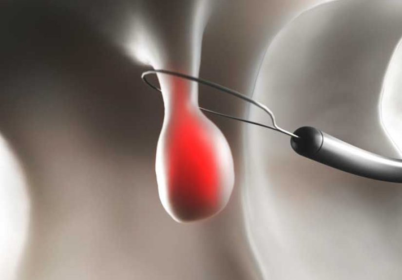
Endoscopic ultrasound (EUS)
EUS is especially valuable when the diagnosis is uncertain or when a doctor needs a closer look at the lesion. It places an ultrasound probe inside the digestive tract, much closer to the pancreas than a standard external ultrasound. That gives clearer images and can help distinguish a pseudocyst from a cystic tumor, walled-off necrosis, or another pancreatic mass.
If needed, EUS can also guide fine-needle aspiration. A small sample of fluid may be collected for analysis when doctors want to rule out another type of pancreatic cyst or evaluate for infection. This is not done for every pseudocyst, but it can be very helpful in the right clinical setting.
Pseudocyst vs. Other Pancreatic Cysts
This is one of the most important parts of diagnosis. Not every cyst near the pancreas is a pseudocyst. Some pancreatic cysts are neoplastic, meaning they arise from abnormal cell growth and may carry cancer risk. That is why doctors pay close attention to whether the person has a history of pancreatitis, what the lesion looks like on imaging, whether there are solid components or nodules, and whether fluid analysis is needed.
A classic pseudocyst usually appears in someone with recent pancreatitis and has imaging features of a fluid collection without the suspicious features more typical of certain pancreatic cystic neoplasms. On the other hand, a “cyst” found in a person with no history of pancreatitis deserves careful evaluation. In that setting, doctors may widen the workup because the diagnosis is not automatically a pseudocyst.
Why Timing Matters in Diagnosis
A pancreatic pseudocyst does not usually appear fully formed overnight. Early fluid collections after acute pancreatitis may evolve, shrink, or organize over time. That is why one imaging study is not always the end of the story. Doctors may recommend repeat imaging to see whether the collection is resolving, stabilizing, or becoming more organized.
This timing issue also helps distinguish a pseudocyst from walled-off necrosis. A pseudocyst contains mostly fluid. Walled-off necrosis contains fluid plus dead tissue and debris. The difference matters because it affects both the diagnosis and what might come next clinically.
When to Seek Medical Care Right Away
Any person with severe abdominal pain, persistent vomiting, fever, jaundice, fainting, confusion, or signs of bleeding needs prompt medical evaluation. That is particularly true if they recently had pancreatitis. A pseudocyst itself may be benign, but its complications are not. Infection, bleeding, rupture, obstruction, and worsening pancreatitis all require urgent attention.
What the Diagnosis Often Means for Patients
For many patients, hearing the word “pancreatic” is immediately terrifying. Understandably so. But a pancreatic pseudocyst is not the same thing as pancreatic cancer, and many pseudocysts improve or resolve without becoming a long-term crisis. The real challenge is figuring out which fluid collections can simply be monitored and which ones are causing symptoms or complications that need more active management.
That is why a careful diagnosis matters so much. It reassures the right patients, identifies the ones who need closer follow-up, and helps avoid confusing a pseudocyst with a very different kind of pancreatic lesion.
Experience Section: What Living Through This Can Feel Like
The experience of a pancreatic pseudocyst often starts long before anyone says the word pseudocyst. For many people, it begins with pancreatitis: a crushing pain in the upper abdomen, nausea that will not quit, vomiting, and a trip to the emergency room that changes the whole week. After the initial hospital stay, a person may assume the worst is over, only to develop persistent fullness, lingering pain, or a strange inability to eat normally. That can be the moment when follow-up imaging reveals a pseudocyst.
One common experience is confusion. A patient may feel “better than before, but not normal.” The fever is gone, yet eating still hurts. The sharp attack has passed, but the upper belly feels swollen, tender, or pressurized. Small meals suddenly feel enormous. Some people describe it as a brick under the ribs. Others say it feels like their stomach has become dramatically less cooperative and far more dramatic.
Another common experience is fear, especially when imaging reports mention a “cystic lesion” near the pancreas. Many patients immediately think of cancer. That anxiety is understandable, and it is one reason doctors work carefully to determine whether the lesion fits the pattern of a pseudocyst or something else. People often remember the waiting period between scans, consultations, and lab results as one of the hardest parts. Physically, they may not be in the worst pain of their lives anymore. Emotionally, however, their imagination is running a marathon.
Patients with chronic pancreatitis may experience pseudocysts differently. Instead of a single dramatic event, they may describe a cycle: pain flare, reduced appetite, weight changes, another scan, another discussion about what the pancreas is doing now. For them, a pseudocyst can feel less like a surprise and more like an unwelcome sequel. Still rude. Still exhausting.
Some people discover a pseudocyst almost by accident. They have imaging after pancreatitis and learn that a fluid collection is still there, even though symptoms are improving. In those cases, the experience can be oddly mixed. There is relief that the finding may be benign, but also frustration that recovery is not as simple as “attack over, life resumes Monday.” Follow-up appointments, repeat scans, diet adjustments, and ongoing symptom watching can make recovery feel longer and more uncertain than expected.
For caregivers and family members, the experience can be stressful too. They may see a loved one who looks weak, eats very little, loses weight, and worries constantly about every new abdominal sensation. Sometimes the most difficult part is not the diagnosis itself but the ambiguity around it: whether the collection will shrink on its own, whether symptoms will settle, and whether the next scan will finally bring clarity.
In real life, a pancreatic pseudocyst is not just a radiology finding. It is often a chapter in a bigger story about pancreatitis, recovery, anxiety, and careful follow-up. That is why clear diagnosis matters so much. It gives patients a name for what is happening, helps separate a pseudocyst from more dangerous look-alikes, and turns a vague, scary problem into something clinicians can evaluate step by step.
Conclusion
A pancreatic pseudocyst is a fluid collection that usually develops after pancreatitis or pancreatic injury. While the name sounds alarming, the condition is often benign and closely tied to inflammation rather than cancer. The biggest clues are the patient’s history, the timing after pancreatitis, the symptoms, and the imaging findings. Many pseudocysts cause abdominal pain, nausea, bloating, or early fullness, while others are found incidentally. Diagnosis usually depends on CT, MRI, ultrasound, or EUS, along with blood tests and careful clinical context.
The bottom line is simple: when a pancreatic fluid collection shows up, accuracy matters more than guesswork. A good diagnosis helps doctors distinguish a pseudocyst from other pancreatic cysts, identify complications early, and guide the next steps with a lot less mystery and a lot more confidence.