Table of Contents >> Show >> Hide
- What Is Proliferative Diabetic Retinopathy (PDR)?
- Why PDR Happens: The Retina’s “Emergency Construction” Plan
- Symptoms and Warning Signs (Including When to Treat It Like a Fire Drill)
- Who’s Most at Risk?
- How Doctors Diagnose PDR
- Treatment Options: What Actually Helps (and Why You Might Need More Than One Thing)
- Screening: The Easiest Win You’ll Ever Get in Eye Care
- Prevention and Day-to-Day Protection: What Actually Moves the Needle
- Frequently Asked Questions
- Real-World Experiences: What the PDR Journey Can Feel Like (About )
- Conclusion
Diabetic retinopathy has a rude sense of timing: it can quietly damage your eyes for years, then show up like,
“Surprise! I redecorated your retina.” The most serious stageproliferative diabetic retinopathy (PDR)
is when the retina starts growing fragile new blood vessels that don’t belong there. These vessels can bleed, scar,
and tug on the retina, raising the risk of major vision loss.
The good news: PDR is treatable, and early detection can be vision-saving. The even better news: you don’t have to
become an eye-science expert to protect your sightyou just need to know what PDR is, why it happens, what symptoms
to respect, and how treatment usually works.
What Is Proliferative Diabetic Retinopathy (PDR)?
Diabetic retinopathy happens when high blood sugar over time damages the tiny blood vessels that feed the retina
(the light-sensitive tissue at the back of your eye). In early stages, those vessels may weaken and leak. In PDR,
the problem escalates: parts of the retina stop getting enough blood, so the eye tries to “fix” the shortage by
growing new vesselsa process called neovascularization.
PDR vs. Nonproliferative Diabetic Retinopathy (NPDR)
You’ll often hear diabetic retinopathy described in two big categories:
-
NPDR (nonproliferative): the earlier stagevessels leak fluid or small amounts of blood, and the retina
can swell. -
PDR (proliferative): the advanced stagenew, abnormal vessels grow and can bleed into the eye, form scar
tissue, and lead to retinal detachment.
And What About Diabetic Macular Edema (DME)?
Here’s the plot twist: many people lose vision from diabetic macular edema (DME) rather than PDR itself.
DME is swelling in the macula (the part of the retina responsible for sharp, central vision). You can have DME with NPDR
or PDR. So if your doctor talks about “PDR and DME,” that’s not a typoit’s two related problems that can overlap.
Why PDR Happens: The Retina’s “Emergency Construction” Plan
Think of your retina like a city that runs on delivery routes. High blood sugar can damage the roads (blood vessels),
causing blockages and poor circulation. When the retina isn’t getting enough oxygen, it releases signalsespecially
vascular endothelial growth factor (VEGF)that tell the body to build new vessels.
Unfortunately, these new vessels are like hastily built scaffolding: thin, leaky, and prone to breaking. They can:
- Bleed into the vitreous (the gel-like fluid inside the eye), causing sudden blur or “floaters.”
- Create scar tissue that can contract and pull the retina out of place.
-
Trigger tractional retinal detachment, where scarring tugs the retina awayan emergency that can threaten
permanent vision. -
Raise eye pressure if abnormal vessels grow in the front of the eye and block fluid drainage, leading to
neovascular glaucoma.
Symptoms and Warning Signs (Including When to Treat It Like a Fire Drill)
One frustrating reality is that diabetic retinopathy can have no symptoms early on. You can have significant
retinal changes and still read street signs like a championuntil you can’t. That’s why eye screening matters.
Common Symptoms People Notice With Advanced Disease
- Blurry vision or fluctuating vision
- Floaters (spots, threads, or “cobwebs” drifting across vision)
- Dark spots or shadows
- Trouble seeing at night
- Sudden vision loss if bleeding is significant
Get Urgent Eye Care If You Notice
- Sudden, significant vision loss in one or both eyes
- A “curtain” or shadow moving across your vision
- New floaters with flashes of light
- Severe eye pain, redness, headache, or nausea (possible high eye pressure)
The goal isn’t to panicit’s to act fast. In PDR, timing can be the difference between “we fixed it” and “we wish we’d seen you sooner.”
Who’s Most at Risk?
Anyone with diabetes can develop PDR, but risk rises with a few common factors:
- Longer duration of diabetes (the “years with diabetes” count matters)
- Higher average blood sugar over time (A1C is a key marker)
- High blood pressure
- High cholesterol
- Kidney disease
- Pregnancy (diabetic retinopathy can worsen during pregnancy)
- Smoking (bad for blood vessels everywhere, including the eyes)
If you’re thinking, “Okay, so basically diabetes plus real life,” you’re not wrong. The key is stacking the odds in your favor with consistent care.
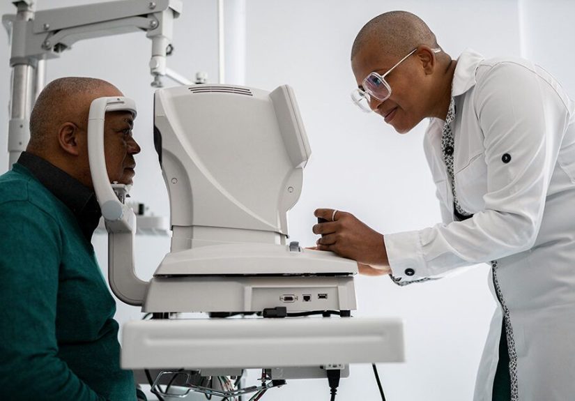
How Doctors Diagnose PDR
PDR is usually diagnosed during a comprehensive dilated eye exam. Eye drops widen the pupils so an eye doctor
can closely examine the retina and optic nerve. It’s painless, though your vision may be blurry and light-sensitive for a few hours afterward
(bring sunglasses and maybe don’t schedule it right before a driving test).
Tests You Might Hear About
- Retinal exam with dilation: looks for abnormal vessels, bleeding, swelling, and scarring.
- Optical coherence tomography (OCT): imaging that helps measure retinal swelling (especially useful for DME).
- Fluorescein angiography: a dye test that highlights blood flow problems and leaking vessels.
- Retinal photography/imaging: sometimes used for screening and monitoring changes over time.
Treatment Options: What Actually Helps (and Why You Might Need More Than One Thing)
PDR treatment focuses on stopping abnormal vessel growth, preventing bleeding and scarring, and protecting the retina.
Your plan depends on how advanced the disease is, whether there’s bleeding, whether DME is present, and how reliably you can follow up.
1) Panretinal Photocoagulation (PRP): “Scatter Laser” Treatment
PRP uses laser spots placed in the peripheral retina to reduce the retina’s demand for oxygen and lower VEGF signals.
In practical terms, PRP helps shrink abnormal blood vessels and lowers the chance of severe vision loss from PDR.
What it’s like:
- Often done in an office or outpatient setting
- May take more than one session
- Some people feel discomfort; numbing drops help
- You may have blurry vision afterward
Trade-offs:
- PRP is durable and doesn’t require frequent injections, which is a big deal if follow-up visits are hard.
- It can reduce peripheral vision or night vision in some peopleyour doctor should talk through that risk.
2) Anti-VEGF Injections: Targeting the “Grow New Vessels” Signal
Anti-VEGF medications are injected into the eye to block VEGF signals, which can help abnormal vessels shrink and reduce leakage.
These injections are widely used for diabetic eye disease (including DME) and are also used for PDR in many situations.
What it’s like:
- Quick in-office procedure with numbing (most people feel pressure more than pain)
- Often started as a series of injections (commonly monthly at first), then spacing out if stable
- Requires reliable follow-upmissing visits can allow PDR to flare again
Practical considerations:
- Injections can be especially helpful if DME is also present, since they treat leakage and swelling too.
- Some people end up using a combination approach: injections plus PRP, depending on how the retina responds.
3) Vitrectomy: Surgery When Bleeding or Scar Tissue Takes Over
If there’s a lot of bleeding into the vitreous (vitreous hemorrhage) or significant scarring and traction, an ophthalmologist may recommend
vitrectomy. This surgery removes the blood-clouded vitreous gel and can address scar tissue pulling on the retina.
It’s often paired with laser treatment during the procedure.
Vitrectomy is usually considered when:
- Bleeding doesn’t clear enough to restore vision or allow laser treatment
- Traction threatens or causes retinal detachment
- Scar tissue is causing major distortion or risk to central vision
4) The “Combo Meal” Approach: PRP + Injections + Surgery (When Needed)
Real life is messy, and so is PDR. It’s common for doctors to combine treatmentsespecially when there’s active neovascularization,
bleeding, and macular swelling in the mix. The best plan is the one that matches your eye findings and your ability to stick with follow-up.
Screening: The Easiest Win You’ll Ever Get in Eye Care
PDR is much easier to manage when caught before major bleeding or detachment. That’s why screening schedules existand why skipping them is a risky sport.
Typical Dilated Eye Exam Timing (Adults)
- Type 1 diabetes: start yearly eye exams within about 5 years of diagnosis (earlier if symptoms or risk factors).
- Type 2 diabetes: get a dilated eye exam at diagnosis, then typically at least yearly.
- Pregnancy with pre-existing diabetes: an eye exam before pregnancy or early in pregnancy, with closer monitoring as recommended.
If exams have been normal and blood sugar is well controlled, some clinicians may space screening out (for example, every 1–2 years).
But once any retinopathy is present, follow-ups are usually at least annualand sometimes much more frequent.
Prevention and Day-to-Day Protection: What Actually Moves the Needle
You can’t time-travel and undo years of high blood sugar (if only). But you can meaningfully lower the risk of progression by focusing on a few key targets:
Keep Blood Sugar in Range
Better long-term glucose control is strongly linked to lower risk of retinopathy progression. A1C is one of the most used tools to measure average blood sugar
over the past few months. Your goal should be individualized with your clinician, but the overall principle is simple: steadier control protects blood vessels.
Manage Blood Pressure and Cholesterol
Retinas don’t like high pressureliterally. High blood pressure and abnormal cholesterol can worsen vessel damage and increase risk of vision problems.
Treating these conditions is not “extra credit”; it’s part of eye care.
Don’t Skip Eye Appointments (Yes, Even When Your Vision Seems Fine)
A large portion of people with diabetes still don’t get recommended yearly eye exams. If you want the simplest actionable step, it’s this:
schedule the dilated exam and keep it.
Know Your Personal “Red Flags”
If you’ve been told you have severe NPDR, early PDR, or DME, ask your eye doctor:
“What symptoms mean I should call the same day?” Then write that answer down. Your future self will thank you.
Frequently Asked Questions
Can PDR be cured?
PDR is treatable, and treatment can stop or slow progression. However, it may not reverse all existing damage. The goal is to preserve vision and prevent
future loss.
Will laser or injections hurt?
Most people tolerate both well. Laser can be uncomfortable, but numbing is used. Injections are typically quick, with numbing drops and a pressure sensation.
Anxiety is commontalk to your doctor about what to expect and ways to make the visit easier.
If my vision is okay, do I still need treatment?
Sometimes, yes. PDR can be “quiet” until it suddenly isn’t. Treatment decisions are based on what the retina looks like, not just what you notice day-to-day.
Real-World Experiences: What the PDR Journey Can Feel Like (About )
If you’ve ever wished medicine came with a “what it’s actually like” trailer, you’re not alone. While every person’s experience is different,
many PDR stories share a few familiar chapters.
Chapter 1: The surprise diagnosis. A lot of people find out they have serious retinal changes during a routine dilated exam
sometimes when they feel totally fine. That can be emotionally confusing: “How can something be wrong if I can still see?”
But PDR often develops before symptoms show up, so the diagnosis can feel like getting a smoke alarm message before you smell smoke.
It’s scary, yesbut it’s also a chance to act early.
Chapter 2: The floaters moment. For others, the first sign is floaterslittle drifting specks or strands.
Some describe it like looking through a snow globe that someone gently shook. If bleeding is heavier, the blur can be dramatic.
The common takeaway people share afterward is simple: don’t “wait it out” just because floaters sometimes fade. Bleeding can recur,
and getting evaluated quickly can prevent bigger problems.
Chapter 3: Laser day (PRP). PRP is one of those treatments where the name sounds like sci-fi, but the reality is more like
a very focused, very boring light show. Some people feel stinging or aching; others mainly feel tired afterward.
It’s common to have temporary blur and light sensitivity. People often say the most annoying part isn’t the procedure
it’s rearranging work schedules and transportation while their vision is dilated or blurry. Planning ahead helps: sunglasses, a ride home,
and a lighter workload that day if possible.
Chapter 4: The injection routine. Anti-VEGF injections can feel intimidating at first (understandably“injection” and “eye”
should not be in the same sentence), but many people report the anticipation is worse than the injection itself.
Visits can become surprisingly routine: check-in, numbing, quick injection, and then back to your day.
The bigger challenge is consistency. People who do well long-term often treat injection visits like a non-negotiable health appointment
the same way you’d treat insulin or blood pressure medication.
Chapter 5: The mindset shift. Many people with PDR describe a turning point where eye care stops being “a once-a-year thing”
and becomes part of their diabetes game plan. That might mean tighter glucose tracking, taking blood pressure seriously,
or finally quitting smoking. Not because anyone loves lifestyle changes, but because vision is personal.
The most common “wish I’d known” is also the simplest: regular eye exams aren’t just about glassesthey’re about protecting your future independence.
Conclusion
Proliferative diabetic retinopathy is serious, but it’s not hopeless. PDR is the stage where abnormal new blood vessels form and can lead to bleeding,
scarring, and retinal detachmentyet modern treatment (PRP laser, anti-VEGF injections, and surgery when needed) can dramatically reduce the risk of severe
vision loss. The best strategy is a two-part plan: keep up with dilated eye exams and manage the drivers that harm blood vesselsblood sugar, blood pressure,
cholesterol, and smoking. If you’ve been diagnosed with PDR, partner closely with a retina specialist, ask questions, and protect your follow-up schedule
like it’s your eyesight’s calendar guardianbecause it is.