Table of Contents >> Show >> Hide
- What subcutaneous emphysema actually is
- How does air get under the skin?
- Common causes (and a few surprising ones)
- Symptoms: what you might notice
- When to treat it like an emergency
- How clinicians diagnose subcutaneous emphysema
- Treatment: what actually helps
- Recovery and what to expect
- Can it be prevented?
- Quick FAQ
- Bottom line
- Experiences people report (and what clinicians often observe)
If you’ve ever pressed on a swollen area and thought, “Why does my skin feel like bubble wrap?”that odd
crackly sensation can be a real medical clue. Subcutaneous emphysema (sometimes called
surgical emphysema) happens when air gets trapped under the skin, usually in the chest,
neck, or face. It can look dramatic, feel strange, and sound even stranger (hello, “Rice Krispies” crunch),
but the bigger question is always why the air got there in the first place.
This guide explains what subcutaneous emphysema is, what causes it, how clinicians diagnose it, what treatment
looks like, and when it’s an emergency. (Spoiler: the air itself is often not the main villainits source is.)
What subcutaneous emphysema actually is
Subcutaneous means “under the skin.” Emphysema in this context means “air where
it shouldn’t be.” So subcutaneous emphysema is simply air (or gas) inside the soft tissues beneath the
skin.
Important clarity break: this is not the same thing as the lung disease called emphysema (a type
of COPD). They share a word, but not a job description. COPD emphysema involves damaged air sacs in the lungs.
Subcutaneous emphysema is air that has wandered into the wrong neighborhood.
The signature sign: crepitus
The classic finding is crepitusa crackling, popping feeling under the fingertips when a clinician
gently palpates the area. People describe it as:
- Crunching snow
- Bubble wrap
- Rice cereal that’s “talking back”
How does air get under the skin?
Air usually enters the subcutaneous tissues because it escapes from somewhere elsemost commonly the
lungs, airways, or chest cavityand then travels along natural tissue planes. Think of it like a
tiny air leak that finds the easiest path and keeps drifting until it hits the soft tissues under the skin.
Subcutaneous emphysema often appears alongside related “air-leak” conditions such as:
- Pneumothorax (air in the pleural space, a.k.a. a collapsed lung)
- Pneumomediastinum (air in the mediastinum, the central chest compartment)
Common causes (and a few surprising ones)
1) Chest trauma and pneumothorax
Blunt or penetrating injurieslike rib fractures, car crashes, sports injuries, or stab woundscan damage the lung
or airways and allow air to escape. A pneumothorax is a frequent partner in crime here, and air can track into the
chest wall and up into the neck.
2) Medical procedures involving the chest or airway
Subcutaneous emphysema can occur after procedures that involve the airway or the pleural space, including:
- Intubation or airway instrumentation
- Mechanical ventilation (especially if airway pressures are high)
- Chest tube placement (or a chest tube that isn’t functioning optimally)
- Thoracic surgery (lung surgery, mediastinal surgery)
- Tracheostomy or neck/airway surgery
This doesn’t mean these procedures are “bad.” It means clinicians watch closely for complicationsbecause air leaks
can happen even when everything is done carefully.
3) Dental work (yes, really)
Some dental procedures use pressurized air or tools that can introduce air into tissuesespecially if there’s a path
through gum tissue or around a tooth. When this happens, people may notice fast facial swelling and crepitus shortly
after treatment. It can be scary (and it can mimic an allergic reaction), so it deserves prompt medical evaluation.
4) Esophageal or gastrointestinal perforation
A tear in the esophagus (including spontaneous rupture after forceful vomiting) can leak air into surrounding
spaces. Subcutaneous emphysemaespecially with neck pain, swallowing pain, fever, or chest paincan be an important
warning sign. Certain bowel perforations can also cause air to dissect upward.
5) Infections that produce gas
Some severe soft-tissue infections can generate gas in tissues. This is uncommon but serious. When subcutaneous air
comes from infection, clinicians look for red flags like fever, rapidly worsening pain, skin discoloration, and
systemic illness.
Symptoms: what you might notice
Symptoms vary depending on how much air is present and what’s causing it. Subcutaneous emphysema itself may cause
discomfort, but the underlying problem (like pneumothorax or airway injury) may cause bigger symptoms.
Common signs and symptoms
- Swelling under the skin (often chest, neck, face)
- Crepitus (crackling sensation when pressed)
- Skin tightness or a puffy appearance
- Voice changes (hoarseness) if air tracks into the neck
- Difficulty swallowing or throat discomfort (varies by cause)
- Chest pain or shortness of breath if a pneumothorax/pneumomediastinum is present
Where it shows up
Most often it’s seen in the chest wall and neck, and it may spread to the face, arms, or even
further. In rare cases, it can track widely because tissue planes act like “highways” for air.
When to treat it like an emergency
Subcutaneous emphysema is not something to “watch and wait” at home if you’re unsure of the cause. Seek urgent
medical careespecially if any of the following occur:
- Trouble breathing, rapid breathing, or feeling like you can’t get enough air
- Chest pain, dizziness, fainting, or blue/gray lips
- Rapidly spreading swelling of the neck/face or difficulty speaking
- Fever, severe pain, or skin changes (possible infection)
- Severe vomiting followed by chest/neck pain (possible esophageal injury)
- Recent trauma (even if it seems “minor”)
The urgency is less about the crackling skin and more about what might be happening underneathlike a pneumothorax,
airway tear, or serious infection.
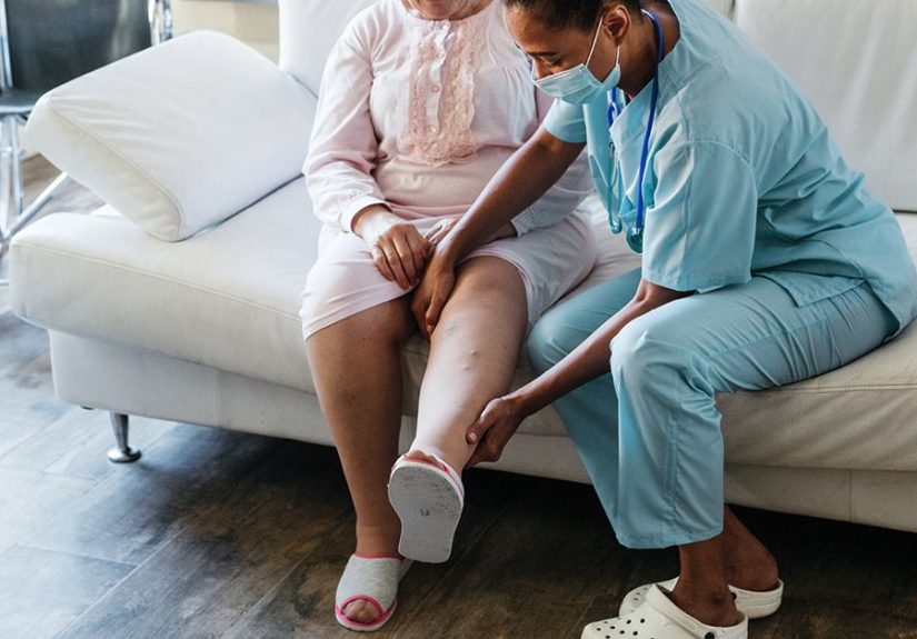
How clinicians diagnose subcutaneous emphysema
1) History and physical exam
Clinicians typically start by asking what happened right before symptoms began: trauma, recent surgery, intubation,
a dental procedure, severe coughing/asthma flare, or vomiting. Then they examine the swollen area and check for
crepitus.
2) Imaging to confirm and find the source
Imaging is often used to confirm air in soft tissues and, crucially, to look for the underlying cause. Common tests:
- Chest X-ray (often a first step)
- CT scan of the chest/neck (more detailed, helps map how far the air has spread)
3) Additional tests (case-dependent)
If clinicians suspect an airway injury, esophageal perforation, or another high-risk cause, they may order targeted
testing (for example, specialized imaging or endoscopic evaluation). The goal is to identify the leak and treat it.
Treatment: what actually helps
Treatment depends on the severity and the cause. Many cases are self-limitedmeaning the body
reabsorbs the air over time once the leak is controlled. But “self-limited” does not mean “self-diagnose.”
Step one: treat the source of the air leak
Examples of source-focused treatment include:
- Pneumothorax: observation for small cases, or a chest tube for larger/unstable cases
- Airway injury: airway stabilization and repair when needed
- Esophageal perforation: urgent hospital management (often surgery/advanced interventions + antibiotics)
- Infection with gas: urgent antibiotics and sometimes surgical management
Supportive care: helping the body reabsorb the air
Clinicians may use supportive strategies such as:
- Oxygen therapy (often used in hospitals; in some cases, higher oxygen can speed reabsorption of gas)
- Pain control and comfort measures
- Monitoring for progression (especially swelling around the neck/airway)
- Adjusting ventilator settings if barotrauma is contributing
Decompression techniques (for severe cases)
If subcutaneous emphysema becomes extensivecausing significant discomfort or threatening breathingclinicians may
consider decompression methods. Depending on the situation, this could include:
- Small subcutaneous drains
- Strategic incisions (“blow holes”) in very select scenarios
- Temporary catheter-based decompression in emergency settings
These are not DIY solutions. Please don’t let the word “blow hole” inspire home improvement energy.
Recovery and what to expect
Once the underlying leak is treated (or stops on its own), the trapped air usually resolves gradually. Mild cases
may improve over days; more extensive cases can take longer, especially if there’s ongoing lung injury or
post-surgical healing.
During recovery, clinicians may mark the borders of swelling, repeat exams, and sometimes repeat imaging to ensure
the air is not expanding and that the underlying issue is improving.
Can it be prevented?
Not alwaysespecially after unexpected trauma. But prevention strategies in clinical settings focus on reducing air
leaks and recognizing them early:
- Careful technique and monitoring during airway procedures
- Appropriate ventilator settings when mechanical ventilation is needed
- Ensuring chest tubes function properly when used
- Post-procedure instructions (for example, avoiding forceful “blowing” behaviors after certain dental work)
Quick FAQ
Is subcutaneous emphysema contagious?
No. It’s air in tissues, not an infection you can “catch.” (Though in rare cases it can be caused by infection.)
Can it happen with COPD?
COPD doesn’t automatically cause subcutaneous emphysema, but people with underlying lung disease may be more
vulnerable to air leaks in certain situations (like severe coughing, barotrauma, or pneumothorax).
Should I massage it out?
You shouldn’t try to treat it at home. The priority is evaluating the cause. Manipulating the tissue won’t fix an
air leak and could delay care if something serious is happening.
Bottom line
Subcutaneous emphysema can look alarming and feel weird, but it’s often a sign rather than the main
problem. The key is to identify where the air is coming fromlungs, airway, chest cavity,
gastrointestinal tract, or (rarely) infectionand treat that source. If you notice sudden swelling with a crackly
sensation under the skin, especially after trauma or a procedure, it’s worth urgent medical evaluation.
Experiences people report (and what clinicians often observe)
The “experience” of subcutaneous emphysema is one of those things that’s hard to forget once you’ve felt it.
People often say the swelling is what they notice firstusually in the neck, chest, or facebecause it changes how
they look in a matter of minutes or hours. A common story goes like this: someone looks in the mirror, sees puffiness
that wasn’t there earlier, and assumes it’s an allergy. Then they touch the area and feel that unmistakable
crackle-pop sensation that doesn’t match any normal swelling they’ve had before.
In emergency and post-op settings, clinicians frequently describe crepitus as “walking on snow” under the fingers.
That tactile clue can be surprisingly helpful, because it pushes the team to think about air-leak problems like
pneumothorax or pneumomediastinum rather than only fluid swelling or inflammation. Nurses and physicians may outline
the borders of the swelling with a marker to track whether it’s expandingbecause change over time matters as much as
the size at any one moment.
People who develop subcutaneous emphysema after a dental procedure often describe a different kind of surprise:
“My tooth was the problemwhy is my face blowing up?” In those cases, the swelling may feel soft and puffy rather
than tight at first, and the crackly feeling can extend along the jawline into the neck. Some people report mild
chest discomfort or a sensation of fullness, which understandably raises anxiety. Clinicians are careful here because
facial swelling could be allergy, hematoma, infection, or aireach one has a different risk profile and treatment.
The presence of crepitus tends to point toward air in tissues.
When subcutaneous emphysema is associated with a pneumothorax, people often talk about the combo of symptoms:
chest pain that feels sharp or worse with breathing, shortness of breath, and then the odd swelling that follows.
Some describe hearing or feeling crackling during movement. In more extensive cases, the swelling can affect the
eyelids or the voice, which can be frighteningespecially if the person feels like they “sound different” or their
throat feels tight. Clinicians take neck and facial involvement seriously because the airway is nearby, even if the
person is breathing comfortably at first.
One of the most consistent emotional experiences people report is uncertainty: “Is this dangerous?” The honest answer
is: it depends on the cause and the trajectory. Many cases improve with monitoring and treatment of the underlying
leak, and the body gradually reabsorbs the air. In the hospital, reassurance often comes from a clear planimaging to
identify the source, repeated exams, and escalation steps if symptoms worsen. People tend to feel better when they
understand that the goal isn’t to “remove every last bubble immediately,” but to stop the leak and make sure breathing
and circulation stay safe while the air fades away.
If you’re supporting a loved one with subcutaneous emphysema, practical comfort helps: keep them calm, encourage slow
breathing if anxiety spikes, follow clinical instructions, and avoid unapproved “fixes” like aggressive massage. The
best experience is the boring onesteady improvement, shrinking borders, and a return to normal without complications.